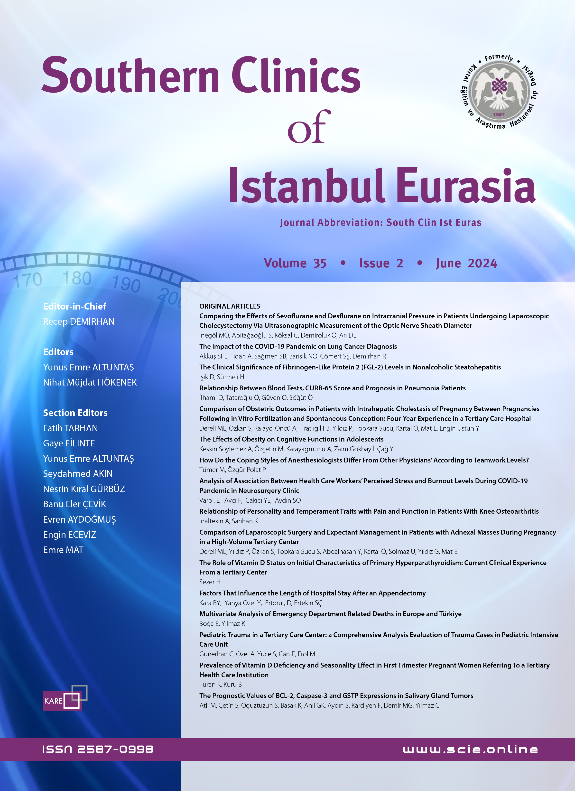
Volume: 32 Issue: 4 - 2021
| RESEARCH ARTICLE | |
| 1. | Management of Children with Severe COVID-19 in a Pediatrics Unit in Istanbul: A Retrospective Study Ayşe Karaaslan, Ceren Çetin, Yasemin Akın, Cem Murat Bal, Recep Demirhan doi: 10.14744/scie.2021.55707 Pages 327 - 332 INTRODUCTION: SARS-CoV-2 is a probable causative agent of severe disease both in children and adults. In this study, we aimed to evaluate the management of hospitalized severe pediatric COVID-19 patients. METHODS: Data on the management of 21 children under the age of 18 who were hospitalized with severe COVID-19 between March 2020 and May 2020 were included in this study. RESULTS: A total of 1109 patients, including 888 outpatients and 221 inpatients, were included in this study. 91 (41.1%) of the 221 hospitalized children were PCR positive for SARS-CoV-2. 21 (23%) of 91 COVID-19 patients were considered severe COVID-19. 10 (47.6%) were females and 11 (52.4%) were males, with a mean±standard deviation (SD) age of 14.4±2.7 years (range; 9 years17.6 years). The most prevalent symptoms at admission were fever (80.9%), cough (76.1%), shortness of breath (23.8%) and myalgia (23.8%). 4 (19%) of 21 patients had underlying diseases. 19 (90.4%) patients were in close contact with confirmed cases in the family. All patients had typical findings on lung computed tomography (CT) and the major CT abnormalities observed were ground-glass opacities. Two patients who needed respiratory support received favipiravir treatment. The mean hospital stay was 7.34±2.65 (516) days. Clinical improvement was achieved in all patients. DISCUSSION AND CONCLUSION: The clinical course of COVID-19 in children is milder and has a better prognosis than adults, but it should be kept in mind that severe cases are defined in the pediatric patient group and these patients should be followed closely. |
| 2. | Prognostic Value of ABO Blood Group in Patients Undergoing Colorectal Cancer Surgery: A Single-Center Experience Aziz Serkan Senger, Selçuk Gülmez, Orhan Uzun, Cem Batuhan Ofluoğlu, Ismail Ege Subasi, Ayhan Öz, Ömer Özduman, Erdal Polat, Mustafa Duman doi: 10.14744/scie.2021.78045 Pages 333 - 337 INTRODUCTION: It has been observed in previous studies that there is a relationship between ABO blood type and gastrointestinal system cancers. However, in studies conducted in different centers, the relationship between the blood type and cancer did not yield the same result. In this single-center study, we investigated the relationship of blood type with colorectal cancer and its effect on prognosis. METHODS: A total of 313 patients who underwent curative surgery for colorectal cancer between January 2013 and December 2019 were included in the study. Data were analyzed retrospectively. Patients with emergency surgery, palliative resection, unavailable records, and distant metastases were excluded from the study. RESULTS: While A blood type was more common in women, other blood types were more common in men. The overall survival for patients with AB blood type was 53.776±7.655 months. And it was significantly worse than the other groups. When examined in the multivariate Cox regression analysis, it was seen that the blood type was effective on the prognosis. DISCUSSION AND CONCLUSION: It was observed that the ABO blood type had an effect on prognosis in colorectal cancer patients. |
| 3. | Analysis of Colonoscopic Diagnosis in Terms of Age, Gender and Symptoms Hakan Uzunoğlu, İsmail Ertuğrul doi: 10.14744/scie.2021.91885 Pages 338 - 344 INTRODUCTION: It was aimed to analyze the relationships between gender, age, and symptom combinations, and the diagnoses determined in colonoscopy. METHODS: Demographic data, symptoms, and colonoscopy results of all patients who underwent colonoscopy in our hospitals endoscopy unit in 2018 were obtained from the hospital records and analyzed. RESULTS: The mean age was 54.5±14.6 years. A total of 66.3% of the patients were over 50 years old. A total of 7.3% of colonoscopy procedures could not be completed. In 168 patients (5.4%) there was fecal occult blood (FOB) positivity, 1900 (60.7%) had rectal macroscopical bleeding, 1893 (60.4%) had constipation, and 1975 (63.1%) had abdominal pain. No abnormal findings were found in 1616 (51.6%) of the patients as a result of colonoscopy, 699 (22.3%) had polyps, 43 (1.4%) had cancer-like masses. The polyp detection rate was significantly higher in patients with positive FOB alone (p=0.003) or patients with rectal bleeding and/or constipation (p=0.001) and significantly lower in patients with constipation (p=0.034). The rates of FOB positivity (p<0.001), polyp (p<0.001), cancer-like mass (p=0.008), and diverticular lesions (p<0.001) were significantly higher in the patients over 50 years of age. The rates of polyps (p<0.001) and diverticular lesions (p<0.001) were significantly higher in FOB-positive patients. DISCUSSION AND CONCLUSION: Findings of the present study showed that patients with FOB positivity or rectal bleeding showed significant pathologies in their colonoscopic examinations, especially over the age of 50, an increase in the rate of the colorectal polyps or cancer-like masses, and a reduced possibility of abnormal findings or malignancies in patients with constipation. |
| 4. | Relationship Between Helicobacter Pylori and Intestinal Metaplasia: A Rural Hospital Experience Ozan Akıncı, Özlem Güngör, Sangar M Faroq Abdulrahman Abdulrahman, Erdem Çomut, Sefa Ergün doi: 10.14744/scie.2020.07088 Pages 345 - 349 INTRODUCTION: Helicobacter pylori (Hp) is a risk factor for gastric cancer, and intestinal metaplasia (IM) is one of the precursor lesions of gastric cancer. The aim of this study was to examine the relationship between Hp and IM. METHODS: A total of 550 patients who underwent upper gastrointestinal endoscopy between October 2018 and December 2019 were included in the study. The patient data of age, sex, endoscopic diagnosis, Hp positivity, IM, atrophic gastritis, and neoplasia were evaluated retrospectively. RESULTS: There were 550 patients enrolled in the study: 228 males (41.5%) and 322 females (59.5%), with a median age of 37.0 years (interquartile range: 18.079.0 years). Hp was detected in 62.7% of the patients, IM in 17.1%, gastric atrophy in 9.1%, and gastric cancer in 2.9%. Among the patients with positive IM, 80 were Hp-positive and 14 were Hp-negative. The rate of IM was significantly higher in Hp positive patients than in Hp-negative patients (p<0.001). In addition, the incidence of Hp-IM coexistence was significantly higher in patients over 50 years of age (p=0.002). DISCUSSION AND CONCLUSION: There was a strong relationship between Hp and IM, and the relationship was correlated with age. Hp eradication is critical to prevent the development of IM, a precursor lesion of intestinal-type gastric cancer. |
| 5. | Our Dartos Pouch Orchiopexy Results Without Transparenchymal Testicular Suture Fixation Yeliz Kart, Canan Öztürk doi: 10.14744/scie.2021.83584 Pages 350 - 353 Objective: To evaluate the operation results in children with the diagnosis of undescended testis who underwent orchiopexy without fixation of transparanchymal sutures. Methods: In this study, 347 patients who were operated on for undescended testis between October 2013January 2020 were retrospectively reviewed. The testis was placed in the dartos pouch and the pouch was narrowed on both sides in patients. Those who underwent surgery with the diagnosis of undescended testis were between the ages of 1 and 15. Transparenchymal suture fixation was not applied in order not to damage testicular parenchymal tissue and adversely affect spermatogenesis. The patients were evaluated in terms of postoperative complications, operation success rate, and recurrence. Results: 347 patients who underwent orchiopexy by narrowing the dartos pouch without transparenchymal suture fixation were evaluated. In the physcial examination performed in the follow-up of 338 patients, it was observed that the testicles were in the scrotum, and the testicular dimensions were normal in the scrotal ultrasonography performed at the 6th month and 1st year postoperatively. 7 patients underwent reoperation due to recurrence, 2 patients underwent orchiectomy for atrophy. Conclusion: In some experimental studies, it was found that the suture passing through the testicular parenchyma damage the testicular parenchyma tissue and affect the germ cell development. For this reason, if the testicle descends into the scrotum without tension, and the dartos pouch is narrowed on both sides without recurrence, testicular parenchymal fixation may not be applied because the suture to be passed through the testicular parenchyma may damage this tissue histologivally. Considering the 97.4% success rate in this study, it was seen that successful orchiopexy could be performed without passing through the testicular parenchyma in suitable patients in order not to adversely affect the spermategenesis. |
| 6. | Evaluation of Pediatric Patients Undergoing Surgical Tracheostomy Nermin Kılıçarslan, Ümran Karaca, Derya Karasu, Seyda Efsun Ozgunay, Mete Kaya doi: 10.14744/scie.2021.35761 Pages 354 - 359 INTRODUCTION: Unlike adult practices, the tracheostomy procedure has higher morbidity and mortality in children. In this study, we aimed to retrospectively investigate our experience of anesthesia in cases who underwent tracheostomy with pediatric surgery. METHODS: Sixty-six children aged 018 years who underwent surgical tracheostomy between 2018-2020 were included. Data included patient demographics, intubation time, tracheostomy indications, anesthetic agents used, and intraoperative and postoperative complications. RESULTS: Of all patients, Thirty-nine (59.1%) were 112 months, 37 (56.1%) were female, and 43 (66.7%) had an underlying neuromuscular disease. Fifty-five patients (84.8%) were hospitalized in the pediatric intensive care unit. The intubation time was 44.16±32.45 days before tracheostomy and the duration of ICU stay was 34.60±26.76 days after tracheostomy. The overall mortality rate was 3.03% (n=2). One of the deaths occurred intraoperatively and the other during the early postoperative period. The most common complications were desaturation in the intraoperative period, bleeding in the early postoperative period, and stoma granulation in the late postoperative period. 63.6% of the patients were discharged. DISCUSSION AND CONCLUSION: We found that prolonged intubation related to neurological disease was the most common indication for tracheostomy in pediatric patients and prolonged tracheostomy opening time did not increase mortality or morbidity. |
| 7. | Epidemiology of burn injuries in Burn Center Osman Esen, Murat Güven, Abdullah Yıldırım, Hamdi Taner Turgut, Çağrı Tiryaki, Murat Burç Yazicioglu, Mustafa Celalettin Haksal, Ali Çiftçi, Hayrünisa Kahraman Esen doi: 10.14744/scie.2021.93270 Pages 360 - 365 INTRODUCTION: The aim of the present study is to determine the epidemiology of burns by assessing demographic features, etiology, findings and burn related factors in burn-injured patients followed-up in a burn center located in the Marmara Region, the most densely populated and industrialized geographical area of Turkey. METHODS: Medical records of 630 in-patients were examined in the burn treatment center. The data relating to the demographic characteristics of patients, burn etiology, percentage of the burned total body surface area, burn degree, burn site, burn agent, place of occurrence, etc. were evaluated. RESULTS: The mean age of 630 in-patients was 27.5±2.18 years. 29.8% of the patients were female, 70.2% were male. The most common causes of burns were respectively; scald (hot water, tea, milk) (37.2%), flame (31.9%), electrical (18.4%) and scald + flame + chemical (11.7%). The average percentage of burned area in the patients was found to be 23.3%.. Inhalation burn was reported in 54 cases (8.9%) and co morbid multiple trauma was reported in 31 cases. DISCUSSION AND CONCLUSION: Burn is a trauma with serious physical and psychological consequences. Although considerable progress has been achieved in burn therapy, attempts for preventing burns and focusing on raising awareness of individuals seem to be more efficient and cheaper. |
| 8. | Are There Any Parameters to Predict the Risk of Screw Breakage Following the Transarticular Screw Fixation in Lisfranc Injuries? A Retrospective Study on 61 Patients Selim Ergün, Mehmet Süleyman Abul, Engin Eceviz doi: 10.14744/scie.2021.58672 Pages 366 - 370 INTRODUCTION: The Lisfranc complex is an osseo-ligamentous structure consisting of multiple tarsal and metatarsal bones, joints, and ligaments. The transarticular screw method is the most commonly preferred fixation technique in patients with Lisfranc injury. However, screw breakage complications can be seen with a considerable frequency. Although there are studies in the literature that associate the risk of screw breakage with the diameter or structure of the screw used, there is still no consensus. The aim of this study is to predict the risk of screw breakage by examining many parameters. METHODS: We retrospectively evaluated 61 patients with lisfranc injuries who underwent transarticular fixation with screws of different diameters and properties. We found screw breakage complications in 9 (14.7%) of the patients. Some demographic and radiological parameters were examined to predict screw breakage in these patients. Age, gender, weight, diameter and structure of the screw used, presence of accompanying cuneiform, cuboid or metatarsal base fracture, how many tarsometatarsal joints were involved and whether anatomical reduction was achieved. In addition, the development of post-traumatic arthrosis during the 2-year follow-up, and the American Orthopedic Foot and Ankle Association (AOFAS) -Middle Foot Score and Visual Analogue Scale (VAS) scores were also compared in clinical evaluations. RESULTS: We could not find any radiological or demographic parameter that could predict the screw breakage complication in the results. We found only that post-traumatic arthritis was significantly more common in patients with screw breakages. We also found that there was no significant difference in AOFAS-midfoot and VAS scores in patients with or without screw breakage complications. DISCUSSION AND CONCLUSION: In the transarticular screw fixation of Lisfranc injury; screws diameter, cannulated or solid structure, the presence of accompanying tarsal or metatarsal injuries and the reduction quality of the injured anatomic structures do not pose any risk factors for the screw breakage complication. |
| 9. | Analysis of the Association Between Minor Cervical Cytological Abnormalities and Consequent Pathology Results According to HPV Types Alper Kahraman, Fırat Tülek doi: 10.14744/scie.2021.52207 Pages 371 - 375 INTRODUCTION: Human papillomavirus (HPV) infection is the leading cause of cervical cancer. Although screening programs involving cervical cytology and HPV DNA have greatly reduced the incidence of cervical cancer in the last decades, attempts to increase the accuracy of these programs are still ongoing. The objective of this study is to evaluate the association of cervical colposcopic biopsy pathology results in women with minor smear abnormalities in an HPV type-specific manner. METHODS: Women who underwent a colposcopic cervical biopsy due to HPV DNA positivity or minor cervical cytological anomalies and tested positive for HPV DNA in a single tertiary center between 2011 and 2019 were retrospectively evaluated. Three groups were formed according to the detected HPV types. The first group consists of women infected with HPV 16 or 18, the second group consists of women infected with only one type of highrisk HPV other than HPV 16/18, and the third group includes patients infected with multiple types of high-risk HPV other than HPV 16/18. RESULTS: Four hundred thirty patients met inclusion criteria within the selected period and were included in the study. The mean age of the population was 33.4±6.6 and the mean parity was 2.1±1.1. The prevalence of ≥CIN 2 lesions in the first and third group of patients were similar, however infection with a single type of high-risk HPV, except for HPV 16/18, was significantly lower in ≥CIN 2 lesions compared to the other two groups. DISCUSSION AND CONCLUSION: Multiple type HPV infections in low grade cytology results even in the absence of HPV 16/18 seems to warrant a cautious approach. |
| 10. | The Relationship Between Vitamin D Levels and Cardiovascular Risk Scores in Geriatric Patients with Type 2 Diabetes Mellitus Damla Ortaboz, Sema Basat, Ridvan Sivritepe doi: 10.14744/scie.2021.25991 Pages 376 - 380 INTRODUCTION: The aim of this study is to evaluate the relationship between vitamin D levels and various cardiovascular risk scoring systems such as QRISK2, BNF, ASSING, SCORE and Framingham in geriatric diabetic patients. METHODS: 60 diabetic geriatric patients with vitamin D deficiency (1030 ng/mL) and 40 geriatric patients with vitamin D deficiency (<10 ng/mL) were included in the study. The scores of the patients indicating cardiovascular disease risks such as QRISK2, BNF, ASSIGN, SCORE and Framingham were calculated. All values were compared between these two groups. RESULTS: While the Framingham risk score (p<0.001), BNF (p=0.001) and SCORE (p<0.001) were found to be significantly higher in patients with vitamin D deficiency, other scores did not significantly differ between the groups. There was a weak but statistically significant negative correlation between 25[OH] D levels with Framingham risk score (p<0.001 r=-0.384), BNF score (p=0.003 r=-0.299), and Score score (p <0.001 r=-0.407). DISCUSSION AND CONCLUSION: In the present study, we found a close relationship between the Framingham, BNF and ASSIGN cardiovascular risk score and serum vitamin D concentrations in diabetic geriatric patients. |
| 11. | The Assessment of Insulin Resistance and Triglyceride/Glucose Index in Nonalcoholic Fatty Liver Disease Melda Çelik, Süleyman Ahbab, Emre Hoca, Hayriye Esra Ataoglu doi: 10.14744/scie.2021.86719 Pages 381 - 387 INTRODUCTION: Insulin resistance is one of the most important risk factors for nonalcoholic fatty liver disease (NAFLD). Homeostasis Model Assessment Insulin Resistance (HOMA-IR) is a marker used to show insulin resistance. Triglyceride/Glucose index (TgG index) is a parameter that can be used to predict NAFLD and is as important as HOMA-IR. In this study, we aimed to determine the value of the HOMA-IR score and TgG index in predicting NAFLD. METHODS: 986 patients who applied to University of Health Sciences Haseki Training and Research Hospital Internal Medicine Clinic between 20172018 and underwent an abdominal ultrasonography scan for any reason were included in the study retrospectively. All medical cases here were investigated in terms of all clinic and laboratory aspects in order to exclude other possible liver-related diseases before they were diagnosed with NAFLD. The patients were categorized and grouped in two different ways. The first is the group with or without NAFLD; the second group was categorized as the control group, prediabetic group and type 2 diabetic group. RESULTS: Our study was conducted with a total of 986 patients, including 470 patients with NAFLD and 516 patients without NAFLD. When the TgG index is calculated; a statistically significant increase was observed in the incidence of NAFLD at levels above 8.4 (p<0.001). The correlation analysis revealed a positive correlation between the TgG index and HOMA-IR (r=0.438). TgG index (p<0.001 OR=3.702), HOMA-IR (p=0.003, OR=1.143), ALT elevation (p=0.001, OR=1.020) were found to be the most effective risk factors when the Backward Stepwise method was used. DISCUSSION AND CONCLUSION: The TgG index was found to be a remarkable predictor-parameter for NAFLD. While HOMA-IR increases the risk of NAFLD by 1.1 times, the the TgG index increases it 3.7 times. In our study, it was also observed that the TgG index increased the risk of NAFLD, independent of HOMA-IR. |
| 12. | Assessment of the Fatigue with Biochemical Data and Corpus Callosum Atrophy in Multiple Sclerosis Patients Tülin Aktürk, Hikmet Sacmacı, Mustafa Erkoç doi: 10.14744/scie.2021.57614 Pages 388 - 394 INTRODUCTION: Fatigue is a common finding in patients with multiple sclerosis (MS) and its etiopathogenesis has not been fully elucidated. The aim of this study was to evaluate the relationship between fatigue and biochemical data and corpus callosum index (CCI) in brain magnetic resonance imaging (MRI). METHODS: In this cross-sectional study, 100 individuals (58 MS patients and 42 control subjects) were included. The disability of the patients was determined with the extended disability status scale (EDSS), and their fatigue was evaluated with the Fatigue Severity Scale (FSS). Biochemical data were analyzed. CCI values were calculated on brain MRI scans. RESULTS: There was no difference between the groups in terms of age, gender, and BMI (p>0.05). In MS patients, folate level and CCI values were found to be statistically significantly lower compared to the control group (p=0.029 and p<0.001, respectively). It was seen that the triglyceride value was significantly higher in the MS group, and there was no significant difference between the groups in terms of other biochemical data. It was found that there was a positive correlation between fatigue and EDSS, and a negative correlation between the CCI and folate value in the MS group. When MS patients with and without fatigue and the control group were compared,only folate levels were found to differ between the groups (p=0.011). DISCUSSION AND CONCLUSION: Our results suggest that fatigue in MS patients is associated with corpus callosum atrophy, a higher level of disability, and lower folate levels. Folate replacement therapy may reduce fatigue, even if folate levels are above the lower limit. Confirming these findings with larger patient series will contribute to a better understanding of fatigue in MS disease and the relationship between fatigue and folate. |
| 13. | The Relationship Between Pain and Vitamin D in Parkinson's Disease İlknur Güçlü Altun, Banu Özen Barut, Anıl Bulut, Nilay Padir, Rahsan Inan doi: 10.14744/scie.2021.74755 Pages 395 - 399 INTRODUCTION: Pain is one of the most common nonmotor symptoms in patients with Parkinson's disease, and sometimes the complaint of refractory pain adversely affects quality of life. The relationship between vitamin D and many types of pain, especially musculoskeletal pain, has been emphasized in different studies. METHODS: The demographic data of 43 idiopathic Parkinson's patients who are above stage 3 according to the Hoehn and Yahr scale, with MMSE 25 and above, and who had not received a diagnosis of dementia, and who had no disease and drug use affecting the vitamin D level, the UPDRS motor subscale, the nonmotor symptom scale and the shortened geriatric depression scale, were recorded. The relationship between vitamin D level and pain was evaluated. RESULTS: Of the patients included in the study, 24 were male and 19 were female. The mean age of the patients was 61.9 (± 9.84) years. The mean disease duration was 3.9 years; the mean UPDRS motor score was 18.3 (± 7.6). According to the nonmotor symptom scale, the patients scored 10.2 (± 5.3) out of 30. 60.5% of the patients had pain complaints. No significant relationship was found between pain and vitamin D level, age, disease duration, UPDRS, drug dose, nonmotor symptom score, GDS, (p>0.05). A significant difference was found between levodopa doses of patients with and without pain in the 60-year-old and younger patients (p = 0.04). Levodopa doses of patients with pain were higher than those without pain. A significant positive correlation was found between the presence of pain and UPDRS in patients over 60 years (p = 0.035). DISCUSSION AND CONCLUSION: As a result of the study, no relation was found between pain and vitamin D levels. In larger patient populations, it was thought to be important to examine the relationship between pain and Vitamin D levels by identifying pain subtypes. |
| 14. | Relationship between Visual Acuity, Refraction and Pachymetry Values, and Topographic Keratoconus Screening Classification Raziye Dönmez Gün, Burak Tanyıldız doi: 10.14744/scie.2021.48379 Pages 400 - 405 INTRODUCTION: To evaluate the relationship between visual acuity, refractive and pachymetric parameters, and topographic keratoconus screening classification (KCSC) in patients without a known diagnosis of keratoconus (KC). METHODS: This retrospective study included 366 eyes of 183 patients for whom topography examination was performed due to clinical suspicion of KC. Visual acuity, refractive and pachymetry parameters, and KCSC result (normal, suspect KC, or KC compatible) according to Sirius topography (CSO, Firenze, Italy) were noted retrospectively. The eyes were divided into 4 groups based on the magnitude of astigmatism (< -1 D, -1 to -2 D, -2 to -4 D, and > -4 D) and the relationship between these groups and topographic KCSC was evaluated. RESULTS: BCVA was higher in the suspect KC group than in the normal group (p=0.008) and lower in the KC compatible group compared to the normal and suspect KC groups (p=0.015, p<0.001, respectively). CCT values were lower in the suspect KC and KC compatible groups than in the normal group (p<0.001) and in the KC compatible group compared to the suspect KC group (p<0.001). Cylindrical values and spherical equivalent were higher in the KC compatible group than the normal and suspect KC groups (p<0.001). In the KC compatible group, fewer eyes had astigmatism of < -1 D or between -1 and -2 D (p<0.05), while significantly more eyes had astigmatism higher than -4 D (p<0.05). DISCUSSION AND CONCLUSION: Patients whose vision did not improve with refraction, who had thin CCT, and/or high astigmatism should undergo topographic examination for KC. Eyes with cylindrical values less than -1 D may be classified as suspect KC or KC compatible, by topography while eyes with astigmatism higher than -4 D are more likely to be classified as KC compatible. |
| 15. | The Incidence of Glaucoma Following Deep Anterior Lamellar Keratoplasty and Penetrating Keratoplasty Ulviye Kivrak, Süleyman Kuğu, Baran Kandemir, Burak Tanyildiz, Hatice Selen Kanar doi: 10.14744/scie.2021.32650 Pages 406 - 411 INTRODUCTION: To evaluate the incidence of glaucoma and the response rates to anti-glaucoma treatment after deep anterior lamellar keratoplasty (DALK) in comparison with penetrating keratoplasty (PK). METHODS: In this retrospective study, 146 patients with DALK and 156 patients with PK were evaluated. All patients were followed up on the 1st, 3rd, 6th, 12th and 24th days after the keratoplasty procedure. The risk factors and incidence of glaucoma and glaucoma treatments were assessed in both the DALK and the PK groups. RESULTS: A total of 156 eyes with DALK procedure and 163 eyes with PK procedure were included. Eleven eyes in the DALK group (7.05%) and 16 eyes in the PK group (9.81%) were diagnosed with glaucoma (p=0. 42). The median number of anti-glaucoma drugs was 1.0 in DALK groups and 1.5 in PK groups (p=0.01). Trabeculectomy with mitomycin C was performed in 4 of the 16 eyes (25%) with glaucoma after the PK (p=0.01). In the DALK group, patients were followed up with mono anti-glaucoma treatment therapy without surgical intervention. DISCUSSION AND CONCLUSION: In our study group, the incidence of glaucoma was 7% in eyes with DALK and 9.8% in eyes with PK. However, the number of glaucoma drugs was significantly higher in eyes with PK procedure than eyes with DALK procedure (p=0.01). Moreover, eyes with DALK have needed less trabeculectomy with mitomycin C compared to eyes with PK. Therefore; our results suggested that the DALK procedure might be more appropriate and safer than PK concerning glaucoma development. |
| 16. | Real-Time Sonoelastography Evaluation of the Lateral Collateral Ligament of Ankle: Comparative Findings Between Athletes and Healthy Subjects Ayşegül Karadayı Büyüközsoy, Tülay Ozer, Nuray Voyvoda, Burak Fariz doi: 10.14744/scie.2021.20591 Pages 412 - 419 INTRODUCTION: The aim of this study is to assess the sonography and elastography findings in the lateral collateral ligament (LCL) injury due to recurrent ankle sprains and to compare them to those of healthy individuals. METHODS: A total of 108 ankles in 54 athletes and 60 ankles in 30 healthy volunteers were included. The LCL, consisting of the anterior talofibular ligament (ATFL), the calcaneofibular ligament (CFL) and the posterior talofibular ligament (PTFL), was evaluated for images in the following three sections: proximal, middle and distal. Longitudinal images of each tendon were obtained using ultrasound (US) and real-time sonoelastography. Degeneration of each tendon was evaluated in four grades and the length and thickness were measured in sonographic images. Real-time sonoelastography images were evaluated in four grades for elasticity pattern and strain ratios. RESULTS: The length of ATFL was shorter in the patient group than in the control group (p=0.028). There was no difference between the patient and control group for ligament thickness. Sonographic grades in the patient group were statistically increased compared to the control group for ATFL, CFL and PTFL ligaments (p<0.001). DISCUSSION AND CONCLUSION: US can be used as a non-invasive diagnostic method to demonstrate ankle LCL injuries in athletes. Real-time sonoelastography provides useful additional information for the evaluation of patients with chronic sprains of the ankle LCL. |
| 17. | The Relationship of REG1A and Ki67 Expression with Prognostic Parameters in Breast Carcinomas Gonca Gül Geçmen, Sevinç Hallac Keser, Dilek İlgici Ece, Sibel Şensu, Ayşegül Selek, Aylin Ege Gül, Ayşe Nimet Karadayı, Nagehan Özdemir Barışık doi: 10.14744/scie.2021.28482 Pages 420 - 426 INTRODUCTION: Invasive breast carcinoma is the most common cancer in women. Literature data on REG1A expression in breast carcinoma was limited. Our study aimed to investigate the relationship between REG1A and Ki67 expressions with prognostic factors that might affect clinical behaviour and disease-free survival in invasive breast carcinomas. METHODS: Patients diagnosed with invasive breast carcinoma at the Dr. Lütfi Kirdar Kartal Training and Research Hospital Pathology Clinic and followed up in the Oncology Clinic between 20072011 were included in our study. Patient records, oncology files, all pathological slides, paraffin blocks were examined are and evaluated. The most appropriate blocks were then selected for immunohistochemical analysis. RESULTS: Our study included 104 cases of primary invasive breast carcinoma. The age of patients ranged between 2495, and the mean age was 53.9±14.8. All cases were female. There was a statistically significant correlation between vascular invasion, histological grade, metastasis, ER, PR, c-erbB2 and REG1A exspression in breast carcinoma. In addition, there was a statistically significant correlation between tumor size, vascular invasion, metastasis, ER, PR, c-erbB2 and Ki67 exspression in breast carcinoma. DISCUSSION AND CONCLUSION: In conclusion, identifying new prognostic parameters, one of which is REG1A, might have a role in the prediction of prognosis and development of new treatment strategies. |
| 18. | Idiopathic Scrotal Calcinosis, Analysis of Epithelial Origin by Immunhistochemical Methods İrfan Öcal, Fazilet Uğur Duman, Zeynep Ayvat Öcal, Fulya Cakalağaoğlu, Mustafa Ozan Horasanlı, Ismail Guzelis doi: 10.14744/scie.2021.89266 Pages 427 - 430 INTRODUCTION: Scrotal calcinosis (SC) is a rare, usually asymptomatic, benign condition characterized by calcium phosphate deposition in the skin of the scrotum. The aim of this study is to investigate the clinical findings of patients diagnosed with scrotal calcinosis and to analyze the presence of possible epithelial origin by immunohistochemistry. METHODS: Fifteen patients diagnosed with scrotal calcinosis by excisional biopsy in two different centers between 2011 and 2020 were included in the study. Demographic, clinical, and laboratory data of the patients were obtained retrospectively from the electronic archive. Hematoxylin & Eosin (H&E) sections prepared from all cases were re-evaluated by two pathologists. Cytokeratin AE1-AE3 immunohistochemical stain was applied to 13 cases for the epithelium of the cyst. RESULTS: The median age of the patients was 32 (468). Patients had ulcer-free, white-yellow, painless nodules measuring 0.5 to 2 cm, mostly in the ventral part of the scrotum, and three had pruritus. Serum calcium levels were normal in eight patients. Histopathological examination showed amorphous and dense, H&E and basophilic calcium deposits with pseudocapsules in the scrotal dermis, most of which had no cyst walls or epithelization. While macrophage infiltration or hyalinization was observed around these areas, ossification was present in one case. While epidermal cysts were observed in the periphery in five cases, calcification, keratinization, and epithelial passage were observed together in two of them. In these areas, the cyst epithelium was positive with cytokeratin AE1-AE3 immunohistochemically. Cysts with cyst epithelium and calcification were smaller than those with only calcification. DISCUSSION AND CONCLUSION: Although the pathogenesis of idiopathic scrotal calcinosis is not clear, the cystic change and calcification in the cyst epithelium and the replacement of the epithelium by calcium appear to be the most likely mechanism after dilatation of the hair follicle. |
| LETTER TO EDITOR | |
| 19. | COVID-19 Infection in a Rare Disease: ROHHAD Syndrome Banu Çevik, Melike Kuvvet Bilen, Büşra Şabano, Elif Bombacı, Kemal Tolga Saracoglu doi: 10.14744/scie.2021.43926 Pages 431 - 432 Abstract | |


