E-ISSN : 2587-1404
ISSN : 2587-0998
ISSN : 2587-0998
Cilt: 33 Sayı: 4 - 2022
| 1. | Front Matter 2022-4 Front Matter 2022-4 Sayfalar I - VIII |
| ARAŞTIRMA MAKALESI | |
| 2. | Pancoast tümörü cerrahisi sonrası üst ekstremite fonksiyonlarının ve egzersiz kapasitesinin değerlendirilmesi Evaluation of Upper Extremity Function and Exercise Capacity after Pancoast Tumor Surgery Talha Doğruyol, Halime Sinem Barutçu, Selime Kahraman, Fatma Tuğba Özlü, Attila Özdemir, Berk Çimenoğlu, Mesut Buz, Fatih Doğu Geyik, Recep Demirhandoi: 10.14744/scie.2022.46793 Sayfalar 341 - 345 GİRİŞ ve AMAÇ: Süperior sulkus tümörlerinin trimodalite tedavisi sonrası kol fonksiyonlarında bozulma gözlenebilmektedir. Bunun sebepleri brakial pleksus ve subklavyen damarların tümör tarafından invazyonu, radyoterapiye sekonder fibrozis, kemoterapiye sekonder nöropati ve cerrahi rezeksiyona bağlı morbidite olarak sayılabilir. Bu çalışmada amaç pancoast tümörü cerrahisi sonrası hastaların kol fonksiyonları ve fonksiyonel egzersiz kapasitelerini değerlendirmektir. YÖNTEM ve GEREÇLER: 2017 Nisan2022 Nisan tarihleri arasında neoadjuvan tedavi sonrası kliniğimizde süperior sulkus tümörü nedeniyle opere edilmiş olan hastalar çalışmaya alındı. Hastalar yaş, cinsiyet, patoloji, neoadjuvan tedavi, rezeksiyon, kullanılan rekonstrüksiyon materyali, ortalama yatış süresi, dren çekilme süresi, drenaj miktarı, morbidite, mortalite, kol fonksiyonları ve fonksiyonel egzersiz kapasiteleri bakımından değerlendirildi. BULGULAR: Belirtilen tarihler arasında kliniğimizde toplamda 18 hastaya pancoast tümörü nedeniyle cerrahi uygulandı. Hastaların tamamı erkekti. Yaş ortalaması 62.7±8.0 olarak hesaplandı. On iki hastaya sağ taraftan girişim, altı hastaya sol taraftan girişim yapıldı. En sık üst lobektomi yapılırken, bir hastaya bilobektomi yapıldı. Morbidite olarak en sık uzamış hava kaçağı (n=5, %27.7) görülürken, cerrahi mortalite görülmedi. Cerrahi olmayan üst ekstremite hareket açıklıkları tam iken, cerrahi olan taraftaki ekstremitede, omuz hareketine göre %2878 arasında eklem limitasyonları saptandı. Hastaların postoperatif 6 dakika yürüme testi ortalaması 536.6±85.7 metre olarak kaydedildi. TARTIŞMA ve SONUÇ: Hastaların kol ve omuz fonksiyonlarının korunması, yaşam kalitelerini arttıracağından daha da önem kazanmıştır. Çalışmamızda pancoast cerrahisi sonrası kol fonksiyonlarının kısıtlandığı ve fonksiyonel egzersiz kapasitesinin düştüğü gösterilmiştir. Bu nedenle bu hastalara preoperatif dönemden itibaren başlayan yoğun fizyoterapi programları planlanması gerektiği kanaatindeyiz. INTRODUCTION: The impairment of arm functions can be observed after trimodality therapy for superior sulcus tumors due to several reasons, such as the tumor invasion of the brachial plexus and subclavian arteries, fibrosis secondary to radiotherapy, neuropathy secondary to chemotherapy, and morbidity caused by surgical resection. This study aimed to evaluate patients arm functions and functional exercise capacities after Pancoast tumor surgery. METHODS: The study included patients that underwent surgery for superior sulcus tumors in our clinic after neoadjuvant therapy between April 2017 and April 2022. The patients were evaluated in terms of age, gender, pathology, neoadjuvant treatment, resection, reconstruction material used, mean hospital stay, drain withdrawal time, amount of drainage, morbidity, mortality, arm functions, and functional exercise capacities. RESULTS: Between the specified dates, 18 patients underwent surgery for Pancoast tumors in our clinic. All the patients were male. The mean age was calculated as 62.7±8.0 years. The operation was performed on the right side in 12 patients and the left in six. The most frequently performed procedure was upper lobectomy, while bilobectomy was performed in one patient. Prolonged air leak was the most common morbidity (n=5, 27.7%), but no surgical mortality was observed. Non-surgical upper extremity ranges of motion were complete, and joint limitations were found in the extremity on the surgical side at a rate varying between 28 and 78%, depending on shoulder motion. The patients mean postoperative six-minute walk test distance was recorded as 536.6±85.7 meters. DISCUSSION AND CONCLUSION: Preserving the arm and shoulder functions of these patients has gained more importance since it increases the quality of life of these patients. Unfortunately, this study showed that the patients arm functions were restricted, and functional exercise capacities decreased after Pancoast tumor surgery. Therefore, we recommend that intensive physiotherapy programs be planned for these patients starting from the pre-operative period. |
| 3. | Acil Servise Üst Gastrointestinal Sistem Kanaması ile Başvuran Hastalarda ROCKALL, AIMS-65 ve GLASGOW BLATCFORD Skorları ile Aktif Kanama Arasındaki İlişkinin İncelenmesi Investigation of the Relationship between ROCKALL, AIMS-65, and GLASGOW BLATCFORD Scores and Active Bleeding in Patients Presenting to the Emergency Department with Upper Gastrointestinal Bleeding Nurhayat Başkaya, Nurdan Yılmaz Şahin, Murat Kekilli, Özge Kibici, Yavuz Katırcıdoi: 10.14744/scie.2022.13540 Sayfalar 346 - 350 GİRİŞ ve AMAÇ: Bu çalışmada, acil departmanına (AD) üst GİS kanaması şüphesiyle başvurup endoskopi yapılan hastalarda, Rockall, AIMS-65 ve Glasgow Blatchford (GBS) skorları ve endoskopide aktif kanama varlığı arasındaki ilişki incelenecektir. YÖNTEM ve GEREÇLER: Çalışma için belirlenen period içerisinde acil servise üst GİS kanama sebebiyle başvuran ve çalışmaya dahil edilen 337 hastanın verileri retrospektif olarak incelendi. Hastaların yaş, cinsiyet, komorbid hastalık, GİS kanama skorları (GBS, Rockall ve AIMS65, endoskopi sonuçları değerlendirildi. BULGULAR: Hastaların %21.3ünde aktif kanama vardı. Aktif kanaması olan hastaların GBS ve Rockall skoru yüksek iken (p<0.05), AIM65 skorunun aktif kanama varlığı ile ilişkisi saptanmadı (p>0.05). GBS için cut-off değeri 11.5, bu değerdeki sensitivite %68.1 spesifite ise %63; Rockall skoru için, cut-off değeri 3.5, bu değerdeki sensitivite %50 spesifite ise %79.6; AIMS65 skoru için cut-off değeri 1.5, sensitivite %36.1 spesifite %74 olarak saptandı. TARTIŞMA ve SONUÇ: Aktif kanama varlığını GBS en iyi gösterirken, bunu Rockall skoru takip etmektedir. AIMS65 skoru aktif kanamayı göstermekte yetersizdir. Aktif kanama varlığını gösterme de bu skorların kullanılabilirliğini doğrulamak için yeni prospektif çalışmalara ihtiyaç vardır. INTRODUCTION: This study aims to examine the association between the Rockall, AIMS-65, and Glasgow Blatchford (GBS) scores to the presence of active bleeding during the endoscopy in patients who are admitted to the emergency department (ED) and suspected of upper gastrointestinal (GI) bleeding. METHODS: The data of 337 patients who visited to the ED due to upper GI bleeding during the period determined for the study were included in the study and analyzed retrospectively. In this context, age, gender, comorbid disease, GIS bleeding scores results (GBS, Rockall and AIMS65, and endoscopy) of the patients were evaluated. RESULTS: Active bleeding has detected in 21.3% of the patients. The GBS and Rockall scores of the patients with active bleeding have found to be high (p<0.05), and there was not an association found between the AIM65 score and the presence of active bleeding (p>0.05). The cutoff value for GBS has determined as 11.5. While the sensitivity at this value was 68.1%, the specificity was 63%. For the Rockall score, the cutoff value has found to be 3.5. While the sensitivity at this value was 50%, the specificity was 79.6%. The cutoff value for the AIMS65 score has found to be 1.5. While the sensitivity at this value was 36.1%, the specificity was 74%. DISCUSSION AND CONCLUSION: The finding that most has been indicated the presence of active bleeding is GBS, followed by the Rockall score. AIMS65 score has been found insufficient for indicating active bleeding. New prospective studies are needed to confirm the usability of these scores in determining the presence of active bleeding. |
| 4. | Çimentosuz Unikondiler Diz Artroplastisi Sonrası Proksimal Tibial Korteksin Kalınlaşma Paterni; Radyolojik Bir Çalışma Remodeling Pattern of the Medial Tibial Metaphysis after a Cementless Unicondylar Knee Replacement; a Radiological Study Enejd Veizi, İzzet Özay Subaşı, Ali Şahin, Hilmi Alkan, Ahmet Firat, Kasım Kılıçarslandoi: 10.14744/scie.2022.09327 Sayfalar 351 - 359 GİRİŞ ve AMAÇ: Unikondiler diz artroplastisi (UDA) öncelikle medial kompartmanın yüzey değişimi için kullanılır ve orta-uzun vadeli sonuçlar umut vericidir. Daha önce birçok çalışmada tibial komponentin etrafındaki kemik dokularda meydana gelen değişiklikler analiz edilmiştir ve bu değişikliklerin, bölgesel olarak artan strese bağlı olduğu bildirilmiştir. Bu çalışmanın amacı, çimentosuz UDA sonrası tibia metafizinin medialinde kemiğin yeniden şekillenme paternini araştırmaktır. YÖNTEM ve GEREÇLER: Bu geriye dönük çalışma Mart 2015 ile Mart 2019 arasında çimentosuz UDA ile tedavi edilen hastalarımız ile yürütüldü. Dahil edilme kriterleri, en az bir olmak üzere, en fazla iki yıl takipli ve yıllık standart ayakta çekilen direkt radyografilerinin mevcut olmasıydı. Toplam 109 hasta dahil edildi. Lateral tibial eminentianın 5 ve 7cm aşağısında iki adet horizontal seviye belirlendi. Bu seviyelerde total metafizier kalınlık ve medial korteks kalınlıkları ölçüldü. Korteks-metafiz oranı (KM) belirlendi ve ölçüm için seri radyografiler kullanıldı. Metafizdeki fokal sklerotik odakların varlığı reaktif üçgen fenomeni olarak değerlendirildi. Hipotezimiz, tüm hastalarda belirli bir düzeye kadar sklerotik değişikliklerin meydana geleceği ve bu değişikliklerin küçük boyuttaki tibial komponentlerde daha fazla olacağı yönündeydi. BULGULAR: KM oranı, ölçülen tüm seviyeler için ameliyat sonrası ilk yıl boyunca azalan bir patern gösterdi. Ameliyat sonrası birinci yılda hastaların %58inde, ikinci yılda ise %80inde tibial komponentin (keel) hemen altındaki metafiz alanında yoğunluk artışı gözlemlendi. Bu yoğunluk artışı grafiye fokal sklerotik bir alan olarak yansıdı ve koronal planda implant taşmaması ile ilişkilendirildi. Tibial komponentin görece büyük olduğu hastalarda KM oranında artış görüldü. TARTIŞMA ve SONUÇ: Unikondiler diz artroplastisi prosedüründen sonra artan kemikteki gerilim stresi, proksimal tibial metafizde kortikal ve kansellöz kemik değişikliklerine yol açar. Bu değişiklikler koronal planda tibial komponentin taşmamasıyla ilişkilidir. INTRODUCTION: Unicondylar knee arthroplasty (UKA) is a surgical procedure primarily used for the resurfacing of the medial compartment and many studies have previously analyzed the changes taking place on the surrounding osseous tissues after the procedure. The purpose of this study is to investigate the effects of bone strain in the medial tibial metaphysis after a cementless unicondylar replacement. METHODS: Patients treated with a cementless UKA between March 2015 and March 2019 was selected for this study. Inclusion criteria were a minimum of 1 and a maximum of 2 years follow-up and presence of standard radiographs of the operated knee at yearly intervals. A total of 109 patients were included in the study. Two lines at a distance of 5 and 7 cm from the lateral tibial eminence were horizontally drawn and the medial cortical thickness and the total cortical distance were measured. A cortex-to-metaphysis (CTM) ratio was established. The increase of density in the metaphysis was analyzed though the reactive triangle phenomenon. We initially hypothesized that some degree of increase in sclerosis would be detected in the medial tibial metaphysis and that the increase would be greater in patients with an implant underhanging, since more cancellous bone would come under strain. RESULTS: The CTM ratio showed a decreasing pattern during the 1st post-operative year for all measured levels. An increase in density at the metaphyseal area just below the keel was observed in 58% of patients during the 1st post-operative year and in 80% during the 2nd year. The increase in the density was correlated with the absence of coronal overhanging. Patients with coronal overhanging of the tibial implant showed an increase in CTM ratio. DISCUSSION AND CONCLUSION: Increased strain after a unicondylar procedure leads to cortical and cancellous bone changes in the proximal tibial metaphysis. These changes depend on the presence or absence of coronal overhanging. |
| 5. | Çocuklarda Preseptal ve Orbital Selülit: Beş Yıllık Tek Merkez Deneyimi Preseptal and Orbital Cellulitis in Children: A Five-Year Single-Center Experience Mehmet Tolga Köle, Ulviye Kıvrak, Yakup Çağ, Serdar Mehmetoğlu, Ayşe Karaaslan, Ceren Çetin, Bilal Yılmaz, Yasemin Akındoi: 10.14744/scie.2022.82574 Sayfalar 360 - 365 GİRİŞ ve AMAÇ: Göz küresi enfeksiyonları çocuk sağlığı ve hastalıkları polikliniklerine nadir başvuru sebeplerinden biri olsa da gelişebilecek intrakranial enfeksiyon, görme kaybı, kavernoz sinüs trombozu gibi ciddi komplikasyonlar nedeniyle hızlı şekilde ayırıcı tanı yapılmalı ve tedaviye başlanmalıdır. Çalışmamız, merkezimizde preseptal ve orbital selülit tanısıyla yatırılarak tedavi edilen çocuk hastaların demografik, klinik, laboratuar ve görüntüleme bulguları ve tedavi yöntemlerinin değerlendirilmesini amaçladı. YÖNTEM ve GEREÇLER: Bu geriye dönük araştırma Ocak 2016 ile Ocak 2021 arasında Kartal Dr. Lütfi Kırdar Şehir Hastanesi, Çocuk Sağlığı ve Hastalıkları ile Göz Hastalıkları Kliniklerinde preseptal ve orbital selülit tanısıyla yatan çocuk hastalarda yapılmıştır. Hastaların klinik ve laboratuvar bulguları karşılaştırılmıştır. BULGULAR: Çalışmaya beş yıl içinde, kriterleri karşılayan 40 hasta (23 erkek, 17 kız) dahil edildi. Hastaların tanı anında ortalama yaşı 145 (6.5181.5) ay idi. Otuz beş hastada (%87.5) preseptal selülit, beş hastada (%12,5) orbital selülit vardı. Tüm vakaların 17sinde (%42.5) altta yatan sebep paranazal sinüzit idi. Yirmi sekiz hasta (tümünün %70i) ampisilin-sulbaktam ve yedi (tümünün %17.5i) hasta seftriakson ile tedavi edilmişti. Orbita enfeksiyonu nedeniyle hastaneye yatırılan 40 hastanın 26sında (%65) hem tanı koymak hem de prognoz takibi için kranial bilgisayarlı tomografi (KBT) çekildi. Ortalama hastanede yatış süresi (gün) preseptal selülit grubunda orbital selülit grubuna göre istatistiksel olarak anlamlı derecede daha düşüktü (p=0.007). TARTIŞMA ve SONUÇ: Çalışmamızda sinüzit, göz küresi enfeksiyonları için en sık predispozan faktör olarak bulundu. Gelişebilecek ciddi komplikasyonlar nedeniyle preseptal ve orbital selülit ayırıcı tanısı hızlı bir şekilde yapılmalı ve hemen tedaviye başlanmalıdır. INTRODUCTION: Although orbital infections are one of the rare reasons for admission to Pediatric Outpatient Clinics, the differential diagnosis should be made and treatment should be started quickly due to the serious complications such as intracranial infection, vision loss, and cavernous sinus thrombosis. Our study aimed to evaluate the demographic, clinical, laboratory, and imaging findings and treatment methods of pediatric patients hospitalized and treated with preseptal and orbital cellulitis diagnosis at our center. METHODS: This retrospective study was conducted on pediatric patients hospitalized with a preseptal and orbital cellulitis diagnosis at Kartal Dr. Lütfi Kırdar City Hospital, Pediatric Outpatient Clinics and Ophthalmology Department between January 2016 and January 2021. The clinical and laboratory findings of the patients were compared. RESULTS: Forty patients (23 boys and 17 girls) who met the criteria over 5 years were included in the study. The mean age of the patients at the time of diagnosis was 145 (6.5181.5) months. Thirty-five patients (87.5%) had preseptal cellulitis and five patients (12.5%) had orbital cellulitis. Paranasal sinusitis was the underlying cause in 17 (42.5%) of all cases. Twenty-eight patients (70%) were treated with ampicillin-sulbactam and seven patients (17.5%) with ceftriaxone. Cranial computerized tomography (CCT) was performed in 26 (65%) of 40 patients hospitalized for orbital infection, for both diagnosis and follow-up prognosis. Mean length of hospital stay (days) was statistically significantly lower in the preseptal cellulitis group than in the orbital cellulitis group (p=0.007). DISCUSSION AND CONCLUSION: In our study, sinusitis was found to be the most common predisposing factor for eyeball infections. Due to the serious complications that can develop, the differential diagnosis of preseptal and orbital cellulitis should be made quickly and treatment should be initiated immediately. |
| 6. | Pulmoner Tromboendarterektomide Anestezi Yönetimi ve Tek Merkez Deneyimlerimiz Anesthesia Management in Pulmonary Thromboendarterectomy and Our Single Center Experience Mustafa Şimşek, Hüseyin Kuplay, Nehir Selcuk, Barış Timur, Türkan Kudsioğlu, Gokcen Orhandoi: 10.14744/scie.2022.99267 Sayfalar 366 - 372 GİRİŞ ve AMAÇ: Bu çalışmada Kasım 2015 ile Aralık 2019 tarihleri arasında merkezimizde pulmoner endarterektomi operasyonu yapılan 26 hastanın sonuçları, ülkemizde sadece belirli merkezlerde yapılan pulmoner tromboendarterektomide anestezi yönetimi ile birlikte değerlendirilerek sunulmuştur. YÖNTEM ve GEREÇLER: Bütün hastalar rutin olarak ileri monitorizasyon yöntemleriyle (5-lead EKG, pulse oksimetre, radial arter kateterizasyonu, ETCO2, santral venöz kateter, serebral oksijenasyon takibi yapmak için rSO2 ve femoral arteryel kateter ve hastaların hemodinamik değerlendirmesini yapmak için (CO, CI, PVR, SVR) termodilüsyon kateteri) monitorize edildi. Rutin olarak bütün hastalar perioperatif TEE ile değerlendirildi. Bütün operasyonlar CPB altında DHCA eşliğinde gerçekleştirildi. Operasyon öncesi ve sonrası PAP, mPAP, CI, CO, PVR, SVR değerleri ölçüldü. Hastaların yoğun bakım kalış süreleri,mekanik ventilatöre bağlanma süreleri, hastanede kalış süreleri, ECLS ihtiyaçları ve mortalite oranları kaydedildi. BULGULAR: Hastaların operasyon sonrası kardiyak index (CI), kardiyak output (CO), oda havasındaki oksijen satürasyonu ve 6 dakikalık yürüyüş mesafesi önemli ölçüde artmıştır. Buna karşılık pulmoner arter basıncında ve pulmoner vasküler rezistansta anlamlı azalmalar tespit edilmiştir. Hastaların mekanik ventilatöre bağlı kalma süresi ortalama 2 (14) gün ve hastanede kalma süreleri ise ortalama 14 (1018) gün olarak tespit edilmiştir. Beş hasta (%19.2) ECLS ye ihtiyaç duymuştur. İlk bir yıl içinde beş hastada (%19.23) mortalite kaydedilmiştir. TARTIŞMA ve SONUÇ: Pulmoner tromboendarterektomi hem cerrahi hem de anestezi yönetimi açısından oldukça komplike bir işlemdir. Bu nedenle ameliyatın başarısı, cerrahi başarının yanı sıra iyi bir perioperatif anestezi yönetimi ile sağlanabilir. INTRODUCTION: In this study, the results of 26 patients who underwent pulmonary endarterectomy in our center between November 2015 and December 2019 are presented by evaluating together with anesthesia management in pulmonary thromboendarterectomy performed only in certain centers in our country. METHODS: All the patients routinely monitored using advanced monitoring methods (5-lead ECG, pulse oximetry, radial artery catheterization, ETCO2, central venous catheter, rSO2 to monitor cerebral oxygenation, femoral artery catheterization, and [cardiac output (CO), cardiac index (CI), Pulmonary vascular resistance (PVR), SVR] thermodilution catheter for hemodynamic evaluation). Routinely, all patients were evaluated with perioperative transesophageal echocardiography. All operations were performed under cardiopulmonary bypass with deep hypothermic circulatory arrest. Pulmonary artery pressure (PAP), mPAP, CI, CO, PVR, and SVR values were measured before and after the operation. The length of stay in the intensive care unit, the duration of mechanical ventilator, the length of stay in the hospital, the extracorporeal life support needs, and mortality rates were recorded. RESULTS: Post-operative CI, CO, oxygen saturation in room air, and 6-min walking distance increased significantly. On the other hand, significant reductions in PAP and PVR were detected. Mean duration of stay on mechanical ventilator was 2 (14) days and average hospital stay was 14 (1018) days. Five patients (19.2%) needed extracorporeal life support. Within the 1st year, 5 mortalities (19.23%) were recorded. DISCUSSION AND CONCLUSION: Pulmonary thromboendarterectomy is a very complicated procedure in terms of both surgery and anesthesia management. Therefore, the success of the operation can be achieved with a good perioperative anesthesia management as well as surgical success. |
| 7. | Hızlı Sıralı Organ Yetmezliği Değerlendirmesi-Mortalite (qSOFAm): Sepsis Hastalarının Mortalitesini Tahmin Etmek İçin Yeni Bir Puanlama Sistemi Quick Sequential Organ Failure Assessment-Mortality (qSOFAm): A New Scoring System to Predict the Mortality of Sepsis Patients Satuk Bugra Yapici, Durdu Mehmet Üzücek, Ahmet Burak Urfalioglu, Dervis Yildiz, Kemal Sener, Adem Kaya, Akkan Avci, Sadiye Yolcudoi: 10.14744/scie.2022.09577 Sayfalar 373 - 378 GİRİŞ ve AMAÇ: Bu çalışmanın amacı, acil servisimize başvuran sepsis hastalarının mortalitesini öngörmede qSOFA'ya dekat ve cinsiyet eklenmesinin tek başına qSOFA'ya üstün olup olmadığını belirlemektir. YÖNTEM ve GEREÇLER: Çalışmamıza 300 sepsis hastasını dahil ettik. Ölüm oranı daha yüksek olan cinsiyet 1 puan daha aldı. Bu değere qSOFA skorunu ve dekatı ekledik ve mortaliteyi tahmin etmek için qSOFA ile yeni skorlama sistemini karşılaştırdık. Ayrıca, mortaliteyi tahmin etmek için yaş için sistolik kan basıncı için bir kesme değeri belirledik. BULGULAR: Hastalarımızın %46'sı kadın, %54'ü erkekti. Çalışma grubunun çoğunluğunu 80'li (%34) ve 90'lı (%26) yaşlarındaki hastalar oluşturdu. Erkeklerde ölüm oranları kadınlara göre daha yüksekti. Mortalite oranı, yaş ve sistolik kan basıncı ile pozitif ilişkiliydi. Yaş ve sistolik kan basıncı için eşik değer 0.71 idi (AUC: 0.799, CI: 0.7500.848, p=0.00). Yaşlarının sistolik kan basıncına oranı 0.71'den (OR: 2.58) yüksek olan hastalarda mortalite riski daha yüksekti. Erkek cinsiyet için qSOFA'ya dekat ve bir puan daha eklendi ve bu değer qSOFA puanı ile karşılaştırıldı. Yeni puanlama sistemi (qSOFA+ dekat+erkek cinsiyet +> 0.71) (CI: -2.49-1.67), mortaliteyi (CI: -1.20-0.80) öngörmede tek başına qSOFA'dan üstündü. TARTIŞMA ve SONUÇ: Yaş/sistolik kan basıncı oranı tek başına mortaliteyi qSOFA'dan daha iyi öngörebilir. qSOFAm skorlama sistemi, sepsis hastalarının mortalitesini belirlemede faydalı olabilir. INTRODUCTION: The aim of this study is to determine whether the addition of decade and gender to quick Sequential Organ Failure Assessment (qSOFA) is superior to qSOFA alone in predicting the mortality of sepsis patients presented to our emergency department. METHODS: We included 300 sepsis patients in our study. The gender with higher mortality received 1 more point. We added the qSOFA score and decade to this value and compared qSOFA and the new scoring system to predict mortality. Furthermore, we determined a cutoff value for age to systolic blood pressure to predict mortality. RESULTS: Forty-six percentages of our patients were female, and 54% were male. Patients in their 80s (34%) and 90s (26%) comprised the majority of the study group. Mortality rates were higher in males when compared with females. Mortality rate was positively related with age and systolic blood pressure. The cutoff value for age and systolic blood pressure was 0.71 (AUC: 0.799, CI: 0.7500.848, p=0.00). Patients were at higher risk for mortality if their ratio of age to systolic blood pressure was higher than 0.71 (OR: 2.58). We added a decade and one more point to the qSOFA for the male gender and compared this value with the qSOFA score. The new scoring system (qSOFA+decade+male gender +>0.71) (CI: −2.49−1.67) was superior to qSOFA alone to predict (CI: −1.20−0.80) mortality. DISCUSSION AND CONCLUSION: Age to systolic blood pressure ratio alone can also predict mortality better than qSOFA. The qSOFAm scoring system may be useful in determining the mortality of sepsis patients. |
| 8. | Sosyal Fobi Komorbidliği olan Bipolar Bozukluk Hastalarında Çocukluk Çağı Travmaları Childhood Traumas in Bipolar Disorder Patients with Social Phobia Comorbidity İsmail Koçdoi: 10.14744/scie.2022.47113 Sayfalar 379 - 383 GİRİŞ ve AMAÇ: Çalışmanın temel amacı sosyal fobi komorbidliği olan bipolar bozukluğu hastalarında çocukluk çağı travmasının etkilerini araştırmaktır. YÖNTEM ve GEREÇLER: Çalışmada, psikiyatrist tarafından Bipolar bozukluk teşhisi almış olan bireyler sırasıyla sosyal fobisi olan (n=39) ve sosyal fobisi olmayan (n=27) olarak, sağlıklı bireyler ise kontrol grubu (n=32) olarak belirlenmiştir. Çalışmada bireylere, sosyodemografik anket, DSM IV Eksen I Bozuklukları için Yapılandırılmış Klinik Görüşme (SCID-I) anketi, Çocukluk çağı travma ölçeği ve Liebowitz Sosyal Anksiyete Ölçeği uygulanmıştır. BULGULAR: Çalışmaya katılan 52 erkek (%53.1) ve 46 kadın (%46.9) bireylerin ortalama yaşları 41.10±10.90 olarak bulunmuştur. Sosyal fobi grubunda 39 birey (%36.7), sosyal fobisi olmayan grupta 27 birey (27.6) ve kontrol grubunda ise 30 birey (%30.6) çalışmada yer almıştır. Sosyal fobisi olan, olmayan ve kontrol grupları arasında Çocukluk Çağı travma anketi açısından istatistiksel açıdan anlamlı bir fark saptanmıştır (p<0.05). Gruplar arasındaki bu farkın hangi gruptan kaynaklandığına ise ikili test ile bakılmış ve bu farklılığın sosyal fobisi olan bipolar hastalarından kaynaklandığı saptanmıştır. Ayrıca, Çocukluk çağı travma anketi, kadın ve erkek cinsiyetleri açısından incelendiğinde kadın bireyler arasında istatistiksel açıdan anlamlı bir fark bulunurken, erkek bireyler arasında ise istatistiksel açıdan herhangi bir anlamlılık bulunmamıştır (p>0.05). TARTIŞMA ve SONUÇ: Çalışmamızda sosyal fobi komorbiditesi olan bipolar bozukluk hastalığının çocukluk çağı travması ile yüksek derece ilişkili olduğu sonucunda varılmıştır. INTRODUCTION: Main purpose of the study is to investigate the effects of childhood traumas on bipolar disorder (BD) patients with social phobia (SP) comorbidity. METHODS: Individuals were divided into three groups, with SP (n=96), without SP (n=27), and without BD divided as the control group (n=32). In this prospective study, the sociodemographic questionnaire, structured clinical interview for DSM-IV axis I disorders (SCID-I), childhood trauma questionnaire, and Liebowitz social anxiety scale were applied to the patients RESULTS: There were 52 (53.1%) men and 46 (46.9) women with the mean age 41.10±10.90 years during the study. Thirty-six patients (36.7%) were in the SP group, 30 patients (30.6%) were in the without SP group, and 32 (32.7) were in the control group. There is a statistically significant difference between bipolar patients with SP, without SP, and the control group in terms of childhood trauma questionnaire (CTQ) scores (p<0.05). It is found that this significant difference originated from the bipolar patients group with SP also, while there is a statistically significant between female patients in terms of CTQ (p<0.05), but there is no statistically significant between male patients in terms of CTQ (p>0.05). DISCUSSION AND CONCLUSION: It was found that SP comorbidity in BD was highly correlated with childhood traumas in our study. |
| 9. | Alt Lomber Faset Eklem Ağrısının Tedavisinde Floroskopi Rehberliğinde Medial Dal Bloğu Sonuçları: 2 Yıllık Takip Results of Fluoroscopy-Guided Medial Branch Block for the Treatment of Lower Lumbar Facet Joint Pain: A 2-year Follow-up Mustafa Umut Etli, Serdar Onur Aydındoi: 10.14744/scie.2022.99907 Sayfalar 384 - 387 GİRİŞ ve AMAÇ: Bu çalışmada, faset eklem bloğu enjeksiyonu adayı olan hastaların iki yıllık takibinde faset eklem bloğu enjeksiyonlarının ağrı kesmedeki yeterliliğini ölçmeyi ve takip sonuçlarını değerlendirmeyi amaçladık. YÖNTEM ve GEREÇLER: Bu çalışmaya 20182020 yılları arasında kliniğimizde faset eklem bloğu enjeksiyonu yapılan 243 hasta dahil edilerek iki yıl boyunca oluşturulan tıbbi kayıtları incelendi. Hastaların demografik özellikleri, ek faset eklem blok enjeksiyonu ihtiyacı, ek cerrahi ihtiyacı, ek cerrahi veya blokaj işleminin nedeni, ilk blokaj ile ek blokaj veya cerrahi arasında geçen süre ve fizik tedavi, algoloji veya ortopedi bölümlerinden ek tedavi ihtiyacı varlığı olup olmadığı değerlendirildi. BULGULAR: Çalışmaya dahil edilen hastaların 93ü erkek, 150si kadındı (ortalama yaş: 54.55, dağılım: 1690). Bunların %62.5i ilk faset blok enjeksiyon girişiminden sonra kalıcı ağrı palyasyonu yaşarken hastaların %5.7sinde ilk işlemden sonra geçici ağrı palyasyonu izlendi ve ortalama 8.4 ay arasında işlemin tekrarlanması gerekti. Hastaların %11.4ü 124 ay arasında dekompresyon ve enstrümantasyon cerrahisi geçirdi. İşlemden fayda görmeyen hastaların tedavileri işlem sonrası fizik tedavi (%14.7), algoloji (%0.8) ve ortopedi (%5.7) bölümlerinde devam etti. TARTIŞMA ve SONUÇ: Faset eklem bloğu enjeksiyonu faset eklemlere bağlı ağrılarda cerrahi yöntemlere göre daha az invaziv olması ve diğer branşlarla uzun süreli tedavi ihtiyacını ortadan kaldırması nedeniyle başarı oranı yüksek bir tedavi yöntemidir. INTRODUCTION: In this study, we aimed to measure the adequacy of facet joint block injections for pain relief during the 2-year follow-up and evaluate the follow-up results of patients who were candidates for facet joint block injection. METHODS: This study included 243 patients who administered facet joint block injections in our clinic between 2018 and 2020. Their medical records created over 2 years were examined. We evaluated the demographic features of patients, the need for an additional facet joint block injection, the need for additional surgery, the reason for the additional surgery or the blockage procedure, and the interval between the first interventional procedure and surgery, as well as additional interventional procedures and the need for additional treatment from the physical therapy, algology, or orthopedics departments. RESULTS: Of the patients included in the study, 93 were male and 150 were female (mean age: 54.55 years, range: 1690 years). Of them, 62.5% experienced pain palliation after the first facet block injection intervention; 5.7% improved after the first procedure, but the procedure had to be repeated between mean 8.4 months; and 11.4% underwent decompression and instrumentation surgery between 1 and 24 months. Those who did not benefit from the procedure continued to receive treatment in the physical therapy department (14.7%), algology department (0.8%), and the orthopedics department (5.7%) after the procedure. DISCUSSION AND CONCLUSION: Facet joint block injection is a treatment method with high a success rates because it is less invasive compared to surgical methods for pain associated with the facet joint and eliminates the need for long-term treatment with other branches. |
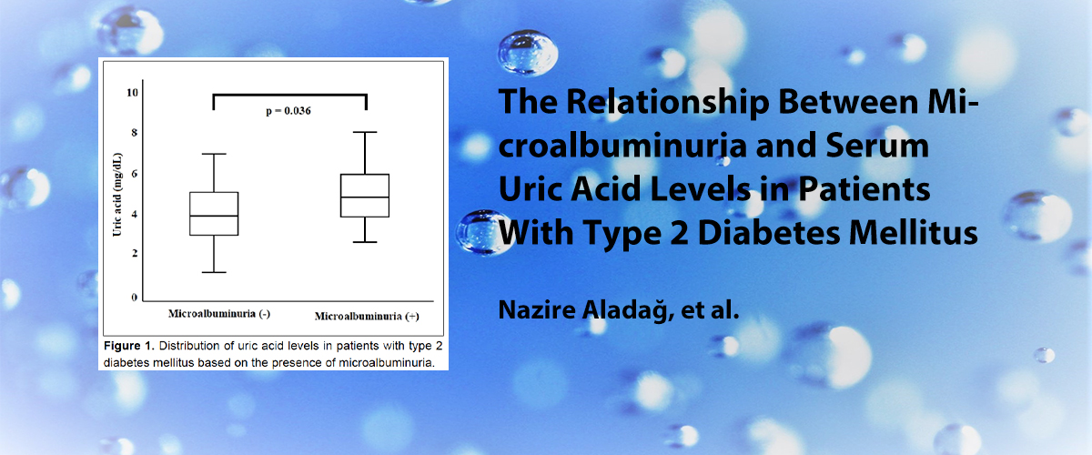
| 10. | Acil Servise Başvuran Akut İskemik İnme Hastalarında Ürik Asit Seviyesinin Mortaliteye Etkisi The Effect of Uric Acid Levels on Mortality in Acute Ischemic Stroke Patients in the Emergency Department Burcu Bayramoğlu, Burcu Genç Yavuz, Şahin Çolak, Dilay Satılmış, Gürkan Akmandoi: 10.14744/scie.2022.36539 Sayfalar 388 - 392 GİRİŞ ve AMAÇ: Ürik asit, serebrovasküler hastalıklar, hipertansiyon ve diyabet üzerine etkisi araştırılmış bir moleküldür. İskemik inme ve ürik asit arasındaki ilişkiyi inceleyen çalışmalarda çelişkili sonuçlar elde edilmiştir. Daha önce yapılan çalışmalarda hem yüksek hem de düşük ürik asit düzeylerinin inme hastalarında kötü prognoz ile ilişkili olduğunu bulmuştur. Bu nedenle, iskemik inmede ürik asit düzeylerinin mortalite, inme şiddeti ve klinik sonlanım üzerindeki etkilerini araştırdık. YÖNTEM ve GEREÇLER: Hastaların demografik özellikleri, kronik hastalıkları, serum ürik asit (SUA) düzeyleri, Ulusal Sağlık Enstitüsü İnme Skalası ve modifiye Rankin Skalası skorları kayıt altına alındı. Hastalar SUA düzeyleri 3.59 mg/dLnin altında, 3.598.5 mg/dL arasında ve 8.5 mg/dLnin üzerindeki hastalar olmak üzere olmak üzere üç gruba ayrıldı. Verilerin karşılaştırılmasında Pearson ki-kare testi kullanıldı. BULGULAR: Çalışmaya %42.4ü kadın olmak üzere 820 hasta dahil edildi. Ortalama yaş 68.53 yıldı. SUA düzeyleri kadınlarda daha düşük bulundu. Orta ve orta-şiddetli inmeler, yüksek mortalite oranı, kötü nörolojik sonlanım ve yoğun bakım ve mekanik ventilasyon ihtiyacı en yaygın olarak SUA düzeyi 8.5 mg/dLnin üzerinde olan hastalarda görüldü, bunu SUA düzeyi 3.59 mg/dLnin altında olan hasta grubu izledi. En düşük mortalite, en iyi nörolojik sonlanım, en az yoğun bakım ve mekanik ventilasyon ihtiyacı ise bu iki SUA düzeyi arasındaki gruptaydı. TARTIŞMA ve SONUÇ: Ürik asit hem oksidan hem de antioksidan özelliklere sahiptir ve etkisi düzey bağımlıdır. En iyi prognoz SUA düzeyi 3.59-8.5 mg/dL arasında olan grupta görüldü. Bu nedenle iskemik inme hastalarını takip ederken ürik asit düzeylerini bu aralıkta tutmak mortalite ve morbiditeyi azaltabilir. INTRODUCTION: Uric acid (UA) is a molecule whose effect on cerebrovascular diseases, hypertension, and diabetes has been investigated. Conflicting results have been obtained in studies examining the relationship between ischemic stroke and UA. The previous studies have found that both high and low UA levels are associated with poor prognosis in stroke patients. Therefore, we investigated the effects of UA levels on mortality, stroke severity, and clinical outcome in ischemic stroke. METHODS: The patient demographics, chronic diseases, serum UA (SUA) levels, National Institutes of Health Stroke Scale scores, and modified Rankin scale (mRS) scores were recorded. The patients were divided into three groups based on SUA levels below 3.59 mg/dL, between 3.59 mg/dL and 8.5 mg/dL, and above 8.5 mg/dL. Pearsons Chi-square test was used to compare the data. RESULTS: A total of 820 patients were included in the study and 42.4% of them were women. The mean age was 68.53 years. SUA levels were lower in women. Moderate and moderate-to-severe strokes, a high mortality rate, poor neurological outcomes, and the need for intensive care and mechanical ventilation were most common in the patients with SUA levels above 8.5 mg/dL followed by the group with SUA levels below 3.59 mg/dL. The lowest mortality, best neurological outcomes, most cases of moderate stroke, and least need for intensive care and mechanical ventilation were in the group with SUA level between 3.59 mg/dL and 8.5 mg/dL. DISCUSSION AND CONCLUSION: UA has both oxidant and antioxidant properties and its effect is level dependent. The best prognosis was seen in the group with SUA levels of 3.598.5 mg/dL. Therefore, maintaining the UA levels within this range when following ischemic stroke patients might reduce mortality and morbidity. |
| 11. | Memenin Paget Hastalığının Moleküler Özellikleri Altta Yatan Duktal Karsinom ile Benzer mi? Üçüncü Basamak Bir Hastaneden Gelen 42 Olgunun Tartışılması How Similar are Molecular Characteristics of Mammary Pagets Disease to Underlying Ductal Carcinoma? Discussion of 42 Cases from a Tertiary Care Hospital Sibel Şensu, Sevinc Hallac Keser, Aylin Ege Gul, Nagehan Ozdemir Barisik, Yesim Saliha Gürbüz, Nusret Erdogandoi: 10.14744/scie.2022.05935 Sayfalar 393 - 398 GİRİŞ ve AMAÇ: Meme Paget hastalığı ve eşlik eden duktal karsinomda östrojen reseptörü, progesteron reseptörü, CerbB2 ekspresyonunu ve moleküler alt tipleri değerlendirmek, bunların uyumunu ve diğer prognostik parametrelerle ilişkisini tartışmak amaçlanmıştır. YÖNTEM ve GEREÇLER: Bu geriye dönük çalışmada Paget hastalığı ve beraberindeki meme karsinomunun klinik ve morfolojik verileri, östrojen/progesteron reseptörü ve CerbB2 immünekspresyonu, moleküler alt gruplar ve sağ kalım değerlendirilmiş olup tüm parametreler istatistiksel olarak karşılaştırılmıştır. BULGULAR: Çalışmaya, memenin Paget hastalığı ve eşlik eden duktal karsinomu olan 42 olgu alınmıştır. Meme örneklerinde 15 olguda (%36) in situ, 4 olguda (%9.5) invaziv ve 23 olguda (%54) in situ+invaziv duktal karsinom saptanmıştır. Aksiller lenf nodu tutulumu 13 olguda (%31) görülmüş olup tümünde invaziv komponent mevcuttur. Östrojen ve progesteron reseptörü ekspresyonu, sırasıyla, duktal karsinomların 16sında (%38) ve 8inde (%19) ve Paget hastalığı olgularının 10unda (%23.8) ve 6sında (%14.2) saptanmıştır. CerbB2 ekspresyonu, duktal karsinomda %93 (39 olgu) ve Paget hastalığında %100 olup %93lük bir uyum göstermiştir. Hem meme duktal karsinomu (%62, 26 olgu) hem de Paget hastalığında (%76, 32 olgu) en sık HER2-baskın moleküler alt tip görülmüş olup %82 uyum saptanmıştır (p=0.03). Exitus grubunda (n=8) sağkalım 46.00 ± 32.64 aydır ve tümünde invaziv duktal komponent mevcuttur (p=0.03). TARTIŞMA ve SONUÇ: Paget hastalığında, eşlik eden duktal karsinoma göre östrojen/progesteron reseptör pozitifliği daha düşük, CerbB2 ekspresyonu ise daha yüksek bulunmuştur. Her iki neoplazide de en belirgin moleküler alt tip HER2-baskın alt tiptir. Tümörlerin hormonal ve CerbB2 immünpozitivitesi prognostik faktörlerle korelasyon göstermezken, invaziv duktal komponentin varlığı sağ kalım ile korelasyon göstermiştir INTRODUCTION: It is aimed to evaluate the expression of estrogen receptor (ER), progesterone receptor (PR), CerbB2 status, and molecular subtypes in mammary Paget disease and concomitant ductal carcinoma and to discuss their concordance and their relation with other prognostic parameters. METHODS: This retrospective study evaluated the clinical and morphological data of the mammary Paget disease and underlying ductal carcinoma; immunohistochemical estrogen/PR and CerbB2 status; molecular subgroups and survival; and statistically compared all parameters. RESULTS: The study included 42 cases of mammary Pagets disease (PD) and concomitant ductal carcinoma. In breast specimens, 15 cases (36%) had in situ, 4 (9.5%) invasive, and 23 (54%) in situ + invasive ductal carcinoma. Axillary nodal involvement was seen in 13 cases (31%) and all had invasive components. Respectively, ER and PR expressions were detected in 16 (38%) and 8 (19%) of the ductal carcinomas and in 10 (23.8%) and 6 (14.2%) of the cases with PD. CerbB2 expression was 93% (39 cases) in ductal carcinoma and 100% in PD with a 93% concordance. The most frequent molecular subtype was HER2-enriched subtype for both mammary ductal carcinoma (62%, 26 cases) and PD (76%, 32 cases) and the concordance was 82% (p=0.03). The survival was 46.00±32.64 months in the exitus group (n=8), all of which had invasive ductal components (p=0.03). DISCUSSION AND CONCLUSION: ER and PR positivity were lower while CerbB2 was higher in Paget disease compared to concomitant ductal carcinoma. The most prominent molecular subtype was HER2-enriched subtype in both neoplasias. While hormonal and CerbB2 status of the tumors did not show any correlation with prognostic factors, existence of an invasive ductal component was the factor that correlated with survival. |
| 12. | Prediyabetli Hastalarda Düşük Doz Metformine Glisemik Yanıt ve Dislipidemi Yanıtı Evaluation of Low Dose Metformin Response and Dyslipidemia Levels in Patients with Pre-diabetes Zeynep Koçdoi: 10.14744/scie.2022.72677 Sayfalar 399 - 405 GİRİŞ ve AMAÇ: Çalışmamızla prediyabetik bireylerde düşük doz metforminin HbA1c ve plazma lipid düzeyleri üzerindeki etkinliğini araştırmayı amaçlanmıştır. YÖNTEM ve GEREÇLER: 20182020 yılları arasında HbA1c düzeyi %5.76.4 arasında olan 357 hastanın dahil edildiği çalışmamızda 1000 mg/gün metformin ile minimum dokuz ay takip edilen olgularda HbA1c düzeyi ve lipid yanıtı değerlendirildi. BULGULAR: HbA1c düzeyleri %66.4 olan grubun %39unda tam yanıt gözlenirken, HbA1c düzeyleri %5.75.9 olan grupta bu %77 yanıt elde edildi. Kırk yaş öncesi elde edilen yanıt %70 iken, 40 yaş üzeri olgularda %36 yanıt elde edilmiştir. Düşük doz metformin kullanan prediyabetli olguların %23ünde nonHDL lipit bileşenlerinde azalma gözlenmiştir. TARTIŞMA ve SONUÇ: Düşük doz metformin, A1cde ve ayrıca nonHDL lipid bileşenlerinde değişen oranlarda azalmaya neden olur. Diabetes mellitusun makrovasküler komplikasyonlarının temeli prediyabet döneminde atılsa da DM ve prediyabetli hastalarda statin ve fenofibrat uygulaması için gereken eşik değerler farklı olup prediyabetikler izole lipid artışı olan olgular gibi tedavi edilebilmektedir. Düşük doz metformin ile HbA1c yanıtının yanı sıra değişen seviyelerde HDL olmayan lipid bileşenlerine yanıt alınabilmektedir. Özellikle prediyabetik dislipidemi grubunda statin ve fibrat tedavisi verilemeyen olgularda kısmi lipit yanıtının olumlu etkileri gözlemlenebilmektedir. INTRODUCTION: This study aimed to investigate the effectiveness of low-dose metformin on the HbA1c and plasma lipid levels in pre-diabetic individuals. METHODS: Between 2018 and 2020, 357 patients with HbA1c levels of 5.76.4% were included in the study. The level of HbA1c and lipid response was evaluated in cases followed up for a minimum of 9 months with 1000 mg/day metformin. RESULTS: A complete response was observed in 39% of the group with HbA1c levels of 66.4%, whereas that in the group with HbA1c levels of 5.75.9% was 77%. While the response obtained before the age of 40 was 70%, the response was obtained in 36% of the cases over the age of 40. A decrease in non-HDL cholesterol components was observed in 23% of the pre-diabetes cases using low-dose metformin. DISCUSSION AND CONCLUSION: Low-dose metformin causes a decrease in A1c, as well as in non-HDL lipid components at varying rates. Although the basis of macrovascular complications of diabetes mellitus is laid during the pre-diabetes, the threshold levels required for the administration of statin and fenofibrate are different in patients with DM and pre-diabetes. Pre-diabetes cases can be treated like cases with isolated lipid increases. In these cases, a response to varying levels of non-HDL lipid components can be obtained in some of the patients, along with an HbA1c response with low-dose metformin. Especially in the pre-diabetic dyslipidemia group, the importance of metformin was observed in the group in which we could not arrange statin and fibrate treatment. |
| 13. | Hiperemezis Gravidarum ve Plasenta Kalınlığı, PAPP-A ve Serbest Beta-HCG ile İlişkisi: Olgu Kontrol Çalışması Hyperemesis Gravidarum and Its Relationship with Placental Thickness, PAPP-A, and Free Beta-HCG: A CaseControl Study Gazi Yıldız, Emre Mat, Didar Kurt, Pınar Yıldız, Gülfem Başol, Elif Cansu Gündoğdu, Betul Kuru, Kasım Turan, Ahmet Kaledoi: 10.14744/scie.2021.93546 Sayfalar 406 - 412 GİRİŞ ve AMAÇ: Çalışmanın amacı, hiperemezis gravidarum ile plasenta kalınlığı, gebelikle ilişkili plazma protein-A ve serbest beta-insan koryonik gonadotropin düzeyleri arasındaki ilişkileri değerlendirmektir. YÖNTEM ve GEREÇLER: Bu çalışmaya 1114. gebelik haftaları arasında kadın hastalıkları ve doğum polikliniğine kombine test için başvuran toplam 263 gebe (93 HGli ve 172 kontrol) dahil edildi. Baş-popo mesafesi (mm) ultrasonografi ile ölçüldü ve gebelikle ilişkili plazma protein-A ve serbest beta insan koryonik gonadotropin değerleri (MoM) laboratuvar sonuçlarından kaydedildi. BULGULAR: Hiperemezis gravidarumlu gebelerin plasenta kalınlığı (p<0.001) ve serbest beta-insan koryonik gonadotropin (p=0.029) değerleri kontrol grubuna göre daha yüksekti. Hiperemezis gravidarum grubunda plasenta kalınlığı, gebelik haftası (p<0.001) ve baş-popo uzunluğu (p<0.001) ile pozitif ve zayıf korelasyon gösterdi. Lineer regresyon analizinde daha yüksek baş-popo mesafesi değerleri ve hiperemezis gravidarum varlığının artmış plasenta kalınlığı (R2=0.159, p<0.001) ile ilişkili olduğu saptandı. TARTIŞMA ve SONUÇ: Hiperemezis gravidarum tanısı konması ve baş-popo mesafesinin artması, artmış plasenta kalınlığı ile ilişkilidir. Bu sonuçla bağlantılı olarak, artmış plasental kalınlık ve serbest beta insan koryonik gonadotropinin de hiperemezis gravidarum için daha yüksek riske neden olduğu görülmektedir. INTRODUCTION: The aim of this study was to evaluate the relationship of hyperemesis gravidarum (HG) with placental thickness, pregnancy-associated plasma protein-A (PAPP-A), and free beta-human chorionic gonadotropin (beta-HCG) levels. METHODS: A total of 263 pregnant women (93 with HG and 172 controls) who applied to the gynecology and obstetrics outpatient clinic for a combined test between 11 and 14 weeks of gestation were included in this study. Crown-rump length (CRL, measured in millimeter) values were measured using ultrasonography, and PAPP-A and free beta-HCG values (MoM) were recorded from laboratory reports. RESULTS: The placental thickness (p<0.001) and free beta-HCG (p=0.029) values of pregnant women with HG were higher than controls. In the HG group, the placental thickness was positively and weakly correlated with gestational week (p<0.001) and CRL (p<0.001). We also found that higher CRL values and the presence of HG were related to increased placental thickness (R2=0.159, p<0.001) by performing linear regression analysis. DISCUSSION AND CONCLUSION: Being diagnosed with HG and having increased CRL is related to increased placental thickness. In relation to this result, increased placental thickness and free beta-HCG also seem to cause a higher risk for HG. |
| 14. | Alt Gastrointestinal Sistem Polipleri: 698 Olgunun Kolonoskopi ve Histopatolojik Özellikleri Lower Gastrointestinal System Polyps: Colonoscopy and Histopathological Features in 698 Cases Yusuf Yavuz, Himmet Durgutdoi: 10.14744/scie.2022.03206 Sayfalar 413 - 416 GİRİŞ ve AMAÇ: Kolon, gastrointestinal sistemde en çok polibin gözlendiği bölgedir. Ülkemizde ve tüm dünyada kolorektal kanserler önemli bir mortalite ve morbidite nedenidir. Submukoza ve mukoza epitelinden kaynaklı bu polipler prekanseröz lezyonlar olabileceği için takip ve tedavisi de önem kazanmaktadır. Bizde bu çalışmamızda ilimizde tespit edilen kolon poliplerinin tür, boyut ve sayı bakımından değerlendirdik ve histopatolojik olarak prekanseröz durumunu inceledik. YÖNTEM ve GEREÇLER: Çalışmamızda ilimizin iki büyük hastanesinde 20132019 yılları arasında yapılan 3654 kolonoskopik inceleme sonrasında tespit edilen, snare ya da forseps yardımı ile polipektomi ya da biyopsi uygulanan 698 kolon polipli vaka değerlendirmeye alındı. Hastaların demografik özellikleri, poliplerin yerleri, polip sayısı, eksize edilen poliplerden en büyük olanlarının boyutları, patolojik tanıları değerlendirmeye alındı. BULGULAR: Çalışmamızda kolon polibi tanısı konan toplam 698 hasta çalışmaya alındı. 698 sayıda hastada toplam 1606 sayıda polibe rastlandı. İşlem başına ortalama polip sayısı 2.3 idi. Çalışmamızda poliplerin boyutlarına göre dağılımı; 527si (%75.5) diminitif polip, 70i (%10) küçük polip, 101i (%14.4) büyük polip olarak saptandı. Polipler distalden proksimale doğru en sık olarak rektumda 278 (%39.8), sigmoid kolonda 175 (%25.1) izlendi. En az polip lokalizasyonu ise 22 (%3.2) ile çekumda görüldü. Poliplerin histopatolojik incelemelerinde en sık tübüler adenom %47 ve hiperplastik polibe rastlandı. 386 (%55) hastada herhangi bir displazi izlenmezken 239 (%34) hastada low grade displazi 6 (%0.9) hastada orta dereceli displazi 67 (%9.6) hastada ise high grade displazi izlendi. TARTIŞMA ve SONUÇ: Kolon kanserlerin gastrointestinal sistemin en sık görülen kanserlerinden olması, kolonda sık olarak bulunan ve yaş ile görülme sıklığı artan kolon poliplerini prekanseröz lezyonlar şeklinde görülebileceğinden dolayı kolonoskopik değerlendirmenin öneminin arttırmaktadır. Endoskopik değerlendirmelerin zamanında yapılması ve tespit edilen poliplerin çıkarılması kanser gelişme riskini düşürme açısından önem kazanmaktadır. INTRODUCTION: Colon is the region where most of the polyps are being observed in the gastrointestinal system. Colorectal cancers are an important cause of mortality and morbidity in our country and all over the world. Since these polyps originating from the submucosa and mucosal epithelium may be precancerous lesions, follow-up and treatment are also important. In our study, we evaluated the type, size, and number of colon polyps detected in our province and examined their histopathological precancerous status. METHODS: In our study, 698 colon polyp cases detected during 3654 colonoscopy examinations performed in the two large hospitals of our city in between 2013 and 2019, and underwent polypectomy or biopsy with the help of snare or forceps, were evaluated. The demographic characteristics of the patients, location of the polyps, number of polyps, sizes of the largest excised polyps, and pathological diagnoses have been evaluated. RESULTS: In our study, a total of 698 patients diagnosed with colon polyps were included in the study. A total of 1606 polyps were detected in 698 patients. The mean number of polyps per procedure was 2.3. In our study, the distribution of polyps according to their sizes was 527 (75.5%) diminutive polyps, 70 (10%) small polyps, and 101 (14.4%) large polyps. Polyps were observed most frequently from distal to proximal as 278 (39.8%) in the rectum and 175 (25.1%) in the sigmoid colon. The least polyp localization was seen in the cecum as 22 (3.2%). In the histopathological examination of polyps, tubular adenoma 47% and hyperplastic polyp were found most frequently. While no dysplasia was observed in 386 (55%) patients, 239 (34%) patients had low-grade dysplasia, 6 (0.9%) patients had moderate dysplasia, and 67 (9.6%) patients had high-grade dysplasia. DISCUSSION AND CONCLUSION: The fact that colon cancers are among the most common cancers of the gastrointestinal system increases the importance of colonoscopy evaluation since colon polyps are found frequently in the colon, the incidence increases by age, and they can be seen as precancerous lesions. Timely endoscopic evaluations and removal of detected polyps gain importance in terms of reducing the risk of cancer development. |
| 15. | Akut Pankreatit Tanılı Hastalarda Serum Lipaz Yüksekliği ile Bilgisayarlı Tomografi Bulguları Arasındaki İlişki The Correlation Between Elevated Serum Lipase Levels and Computed Tomography Findings in the Patients with Acute Pancreatitis Rasime Pelin Kavak, Nezih Kavak, Nurcan Ertan, İlkay Güler, Nurgül Balci, Ahmet Sekidoi: 10.14744/scie.2022.88709 Sayfalar 417 - 422 GİRİŞ ve AMAÇ: Amacımız akut pankreatit tanılı (AP) hastalarda serum lipaz yüksekliği ile bilgisayarlı tomografi (BT) bulguları arasındaki ilişkiyi değerlendirmektir. YÖNTEM ve GEREÇLER: Acil serviste AP tanısı alan hastalar serum lipaz değerlerine göre, normal sınırın üç katı (grup 1) ve on katı (grup 2) olarak iki gruba ayrıldı. Gruplar arasında demografik özellikleri (yaş, cinsiyet), karın ağrısnın vasfı (tipik, atipik), başvuru süresi ve BT bulgularının var ve yok olması açısından karşılaştırıldı BULGULAR: Yüz yirmi iki hastanın %53.3ü kadın idi. Hastaların yaş ortalaması 62.17±6.74 (min 35-max 75) yıl idi. Hastaların %63.1i grup 2de yer almaktaydı. Hastaların başvuru süresi ortalaması 14.42±10.11 (min 4-max72) saat idi. %63.9 hastada karın ağrısı atipik vasıfta idi. Hastaların %56.6sında BT bulguları mevcuttu. Hastaların %3.7sinde pankreas nekrozu saptandı. Gruplar arasında BT bulgularının var ve yok olması açısından farklılıklar saptandı (p<0.05). Grup 2de BT bulgularının var olma oranı daha fazla idi. BT bulguları olan ve olmayan hastalar arasında karın ağrısı tipik/atipik vasıfta olması oranları arasında fark saptandı (p<0,001). BT bulguları var olan hastalarda karın ağrısının atipik vasıfta olma oranı daha yüksek idi (p<0.001). TARTIŞMA ve SONUÇ: AP tanılı hastalarda serum lipaz değeri arttıkça BT bulgularının var olma olasılığıda artmaktadır. INTRODUCTION: This study was evaluation of the relationship between serum lipase elevation and computed tomography (CT) findings in patients with acute pancreatitis (AP). METHODS: Patients who received AP diagnosis in the emergency department were divided into two groups according to their serum lipase values that were three (group 1) and 10 times (group 2) higher than the normal upper limit, respectively. Demographic characteristics (age and gender), nature of abdominal pain (typical and atypical), duration of presentation, and CT findings were compared between groups in terms of present and absent. RESULTS: About 53.3% of 122 cases were female. The mean value of patient age in the study was 62.17±6.74 (min 35max 75) years. About 63.1% of the patients were in Group 2. Themean ED admission interval of the patients was 14.42±10.11 (min 4max 72) h. The nature of abdominal pain was atypical in 63.9% of the patients. CT findings were present in 56.6% of the patients. Pancreatic necrosis was detected in 3.7% of the patients. Dissimilarities between the two groups were identified in respect of the presence or absence of CT findings (p<0.05). The present rate of CT findings was greater in Group 2. Furthermore, the rates of typical/atypical nature of abdominal pain between patients whose CT findings were present and absent had significant distinction (p<0.001). The rate of atypical nature of abdominal pain was higher in patients with present CT findings (p<0.001). DISCUSSION AND CONCLUSION: As the serum lipase value increases in patients with AP, the probability of CT findings being present increases. |
| 16. | Safra Kesesi Displazi ve Kanserlerinde Apopitoz ve Çoklu İlaç Direnci İlişkili Belirteçlerin İmmünohistokimyasal Olarak Değerlendirilmesi Immunohistochemical Evaluation of Apoptosis and Multidrug Resistance-Related Markers in Gallbladder Dysplasia and Carcinoma Kayhan Başak, Derya Demir, Arzu Kaya Koçdoğan, Serpil Oğuztüzündoi: 10.14744/scie.2022.82712 Sayfalar 423 - 428 GİRİŞ ve AMAÇ: En agresiv seyir gösteren, kötü prognoza sahip ve tedavi direnci göstreme eğiliminde olan tümörlerden biri olan safra kesesi karsinomlarında (SKK) tedavi başarısı arayışı günümüzde devam etmektedir. Solid tümörlerin çoğunda saptanan genetik değişikliklerle ilgili yolakları hedef alan tedaviler bu tümörlerin tedavisinde yeni ümit olmaktadır. Bu tedavi modalitelerinden bazıları apopitoz ilişkili yolakları hedeflemekte olup, mTOR, p38, Bcl-2 ve caspase-3 bu yolağın önemli bileşenlerindendir. YÖNTEM ve GEREÇLER: Çalışmada 27 SKK ve 62 safra kesesi displazisi tanısı olan olguların parafin bloklarına mTOR, caspase-3, p38, Bcl-2, LL- 37, MDR1, MRP1, MRP6, and MRP7 immünohistokimyasal boyaması uygulandı. İmmünohistokimyasal olarak boyanmış kesitler değerlendirildi ve skorlandı. BULGULAR: mTOR, p38 ve caspase-3 ekspresyonları displazi ve kanser gruplarında, displastik ve malign hücrelerde istatistiksel olarak anlamlı artmış bulundu. MRP1 ve MRP7 nin ekspresyonlarında anlamlı farklılık yokken, MRP6 anlamlı olarak fazla eksprese ediliyordu. TARTIŞMA ve SONUÇ: Bu çalışmada safra kesesi displastik ve malign hücrelerinde mTOR, p38 ve caspase-3 ekspresyonunun artması, safra kesesinde karsinogenez sürecinde rolü olduğunu gösterebilir. Çalışma ayrıca MRP6nın safra kesesi karsinomunda ilaç direncinin gelişmesinde rol oynayabileceğini de göstermektedir. INTRODUCTION: The search for treatment success in gallbladder carcinomas, which is one of the tumors with the most aggressive course, poor prognosis, and tendency to show resistance to treatment, continues today. Treatments targeting pathways related to genetic changes detected in most solid tumors offer new hope in the treatment of these tumors. Some of these treatment modalities target apoptosis-related pathways, and mammalian target of rapamycin (mTOR), p38, Bcl-2, and caspase-3 are important components of this pathway. METHODS: In the study, mTOR, caspase-3, p38, Bcl-2, LL-37, MDR1, multidrug resistance protein (MRP)1, MRP6, and MRP7 immunohistochemical staining were applied to paraffin blocks of 27 gallbladder cancer and 62 cases with gallbladder dysplasia. The immunohistochemically stained sections were evaluated and scored. RESULTS: mTOR, p38, and caspase-3 expressions were found to be significantly increased in dysplasia and tumor groups, and in dysplastic and malignant cells. While there was no significant difference in the expression of MRP1 and MRP7, MRP6 was significantly overexpressed. DISCUSSION AND CONCLUSION: In this study, increased expression of mTOR, p38, and caspase-3 in the dysplastic and malignant cells of the gallbladder may show that it has a role in the carcinogenesis process in the gallbladder. The study also shows that MRP6 may also play a role in the development of drug resistance in gallbladder carcinoma. |
| 17. | D Vitamini Eksikliğine Bağlı Hipokalsemik Nöbetleri Olan Çocuklarda Nörolojik Prognozun Değerlendirilmesi Evaluation of Neurological Prognosis in Children with Hypocalcemic Seizures Due to Vitamin D Deficiency Gül Demet Kaya Özcora, Elif Söbü, Türkan Uygur Şahindoi: 10.14744/scie.2022.67299 Sayfalar 429 - 434 GİRİŞ ve AMAÇ: Çalışmamızda hipokalsemik nöbet tanısı alan süt çocuklarının bir yıllık izlemdeki nörolojik bulgularının değerlendirilmesi amaçlandı. YÖNTEM ve GEREÇLER: Çalışmaya Temmuz 2017Temmuz 2018 tarihleri arasında hipokalsemik nöbet tanısıyla izlenen, yaşları 6 gün ile 24 ay arasında 32 olgu dahil edildi. Kaba motor, ince motor, dil ve sosyal gelişim düzeyleri tanı anında ve altıncı ayda Denver gelişim testi ile değerlendirildi. BULGULAR: Olguların %72si erkek (n=23) ve %28i (n=9) kızdı. Düzeltilmiş serum kalsiyum seviyesi ortalama 6.03±1.06 mg/dL, fosfor 5.2±1.72 mg/dL, alkalen fosfataz 575.2±405.8 U/L; parathormon 231.4±123.8 ng/ml ve 25-OH vitamin D düzeyi 9.6±8.5 ng/ml olarak bulundu. Annelerin D vitamini düzeyleri incelendiğinde ortalama 7.16±2.62 ng/ml saptandı. Kaba motor gelişimdeki altı aylık fark istatistiksel olarak anlamlı bulundu (p<0.001), ince motor gelişimdeki altı aylık fark istatistiksel olarak anlamlıydı (p <0.001). Sosyal gelişim ve dil gelişiminin değerlendirilmesindeki altı aylık fark istatistiksel olarak anlamlı değildi (sırasıyla p=0.083 ve p=0.180). Altı aylık süreçte hastaların ince ve kaba motor gelişimdeki fark istatistiksel olarak anlamlı kabul edildi (p<0.001). TARTIŞMA ve SONUÇ: D vitamini bir nörohormondur, nörotransmiter üretimi, salımı ve geri alımında rol oynayan ve beyin hücresi çoğalması, farklılaşması ve gelişimi aşamalarında önemli rol alan kalsiyum metabolizmasının önemli bir düzenleyicisi olduğu için eksikliğinde psikomotor gerilikte görülebilmektedir. INTRODUCTION: This study aimed to evaluate the neurologic findings of patients who had hypocalcemic seizures due to vitamin D deficiency during a 1 year period. METHODS: Thirty-two patients aged between 6 days and 24 months who were followed up for hypocalcemic seizures between July 2017 and July 2018 were included in the study. Gross motor, fine motor, language, and social developmental levels were evaluated using the Denver developmental test at the time of diagnosis and the 6th month. RESULTS: Seventy-two percentages of the patients were male (n=23) and 28% (n=9) were female. Corrected serum calcium level averaged 6.03±1.06 mg/dL, and phosphorus was 5.2±1.72 mg/dL, alkaline phosphatase was 575.2±405.8 U/L, parathormone was 231.4±123.8 ng/mL, and 25 OH-vitamin D levels were found as 9.6±8.5 ng/mL. When the vitamin D levels of the mothers were examined, the average was 7.16±2.62 ng/mL. The 6-month difference in both gross and fine motor development was found to be statistically significant (p<0.001 and p<0.001, respectively). The 6-month difference in the evaluation of social development and language development was not statistically significant (p=0.083 and p=0.180, respectively). The 6-month difference in fine and gross motor development over the patients was considered to be statistically significant (p<0.001). DISCUSSION AND CONCLUSION: Psychomotor retardation is also observed in infants with vitamin D deficiency, because vitamin D is a neurohormone that has an important role in calcium metabolism, which plays a role in neurotransmitter production, release, reuptake, and in stages of brain cell proliferation, differentiation, and development. |
| 18. | Apendektomi Spesimenlerinde Saptanan Apendiks Tümörlerinin Analizi: Tek Merkezli Deneyim Analysis of Appendiceal Tumors Detected in Appendectomy Specimens: Single Center Experience Ahmet Başkent, Murat Alkan, Mehmet Furkan Başkentdoi: 10.14744/scie.2022.52385 Sayfalar 435 - 441 GİRİŞ ve AMAÇ: Bu çalışmanın amacı, merkezimizde apendektomi spesimenlerinde saptanan apendiks tümörlerini belirlemek ve bu tümörlerin insidansı ile klinikopatolojik özelliklerini analiz etmektir. YÖNTEM ve GEREÇLER: Ocak 2015Aralık 2021 tarihleri arasında hastanemizde yapılan toplam 6110 apendektomi olgusu geriye dönük olarak değerlendirildi. Bu olguların demografik özellikleri ile histopatolojik incelemeleri analiz edildi. Apendiks tümörü (AT) saptanan olguların yaşı, cinsiyeti gibi demografik özellikleri ile ameliyat prosedürleri ve histopatolojik sonuçları incelendi. BULGULAR: 6110 apendektomi örneğinin histopatolojik incelemesinde toplam 44 (%0.72) AT saptandı. Bunlar temel olarak ikiye ayrıldı. Birincisi apendiksin nöroendokrin tümörleri (ANET) 33 (%75) olgu ve ikincisi de 11 (%25) olgu ile apendiksin non-karsinoid tümörleri (ANCT) yani epiteliyal tümörleridir. ANCT yani epiteliyal tümörleri 6 (%54.5) olguda düşük dereceli müsinöz neoplazm (LAMN), 2 (%18.2) olguda müsinöz komponentli adenokarsinom, 2 (%18.2) olguda apendikse metastaz yapmış (akut apandisit kliniği ile opere edilen) adenokarsinom ve 1 (%9.1) olguda da ANET komponentli adenokarsinom saptandı. ANETlerden 26 (%78.8) olguya sadece appendektomi yapılırken 7 (%21.2) olguya sekonder olarak SH yapıldı. ANCTlerdeki 6 (%54.5) olguya sadece apendektomi, 2 (%18.2) olguya peroperatif apendektomi ile birlikte geniş lokal eksizyon, 3 (%27.3) olguya da sekonder olarak sağ hemikolektomi (SH) yapıldı. 44 AT içinden toplamda 10 (%22.7) olguya sekonder sağ hemikolektomi yapılmış oldu. Sekonder SH yapılan 2 (%4.5) olguda rezidü tümör saptandı. Rezidü tümör saptanan olgulardan biri ANET Grade 3, diğeri müsinöz komponentli adenokarsinom olup cerrahi sınırlar temiz olarak raporlandı. Apendikse metastaz yapan iki hasta ile apendiks müsinöz komponentli adenokarsinomu olan bir hasta, yani toplamda 3 (%6.8) hasta hayatını kaybetti. Çalışmamızda saptanan müsinöz adenomlu 4 olgu çalışmaya dahil edilmedi. TARTIŞMA ve SONUÇ: Apendektomi materyallerinde malignite olasılığı nadirdir ve genellikle apendektomi sonrası patolojik incelemelerde tesadüfen saptanır. Bu sebeple bütün apendektomi örneklerinin rutin olarak histopatolojik inceleme için gönderilmesini öneririz. Karsinomlar, ANETlere göre daha kötü bir prognoza sahiptir. İleri evre ANET ve apendiks epiteloid tümörlerinde tamamlayıcı sağ hemikolektomi önerilmektedir. INTRODUCTION: The aim of this study is to identify appendiceal tumors (AT) detected in appendectomy specimens in our center and to analyze the incidence and clinicopathological features of these tumors. METHODS: A total of 6110 appendectomies performed in our hospital between January 2015 and December 2021 were evaluated retrospectively. Demographic characteristics and histopathological examinations of these cases were analyzed. Demographic characteristics such as age, gender, surgical procedures and histopathological results of cases with AT were analyzed. RESULTS: A total of 44 (0.72%) AT were detected in the histopathological examination of 6110 appendectomy specimens. These are basically divided into two. The first is appendiceal neuroendocrine tumors (ANET) with 33(75%) cases and the second is appendiceal non carcinoid tumors (ANCT), that is, epithelial tumors, with 11 (25%) cases. ANCTs, that is, epithelial tumors, were detected in the following four features: Low-grade appendiceal mucinous neoplasm in six cases (54.5%), adenocarcinoma with mucinous component in two cases (18.2%), adenocarcinoma with metastasis to the appendix (operated with acute appendicitis clinic) in two cases (18.2%), and adenocarcinoma with ANET component in one case (9.1%). Only appendectomy was performed in 26 (78.8%) cases in ANETs, while secondary right hemicolectomy (RH) was performed in 7 (21.2%) cases. In ANCTs, only appendectomy was performed in 6 (54.5%) cases, wide local excision with perioperative appendectomy in 2 (18.2%), and secondary RH in 3 (27.3%) cases. Secondary RH was performed in 10 (22.7%) cases out of 44 AT patients. Two patients who metastasized to the appendix and one patient with appendiceal carcinoma, that is, 3 (0.05%) patients in total, died. DISCUSSION AND CONCLUSION: The possibility of malignancy in appendectomy materials is rare and is usually detected incidentally in pathological examinations after appendectomy. Therefore, it is recommended that all appendectomy specimens be routinely sent for histopathological examination. Carcinomas have a worse prognosis than ANETs. Complementary RH is recommended for advanced ANET and appendiceal epithelioid tumors. |
| 19. | Küçük Hücreli Dışı Akciğer Kanserinin Histolojik Alt Tipleri ile Sağkalım Arasındaki İlişki: 1887 Hastanın Geriye Dönük Kohort Analizi Association between Histologic Subtypes of Non-Small Cell Lung Cancer and Survival: Retrospective Cohort Analysis of 1887 Patients Mesut Buz, Seyyit Dincerdoi: 10.14744/scie.2022.59862 Sayfalar 442 - 447 GİRİŞ ve AMAÇ: Küçük hücreli dışı akciğer kanserinin histolojik alt tipleri hastalığın sonucu ile ilişkili olabilir. Nadir fakat agresif bir tümör olan adenoskuamöz karsinom, zayıf sağkalım oranları ile ilişkilendirilmiştir. Bu çalışmada küçük hücreli dışı akciğer kanserinin histolojik alt tiplerine sahip hastaların cerrahi tedavi sonrası sağ kalım oranlarının değerlendirilmesi amaçlanmıştır. YÖNTEM ve GEREÇLER: 20002009 yılları arasında küçük hücreli dışı akciğer kanserli hastalarda cerrahi tedavi uygulanan tek merkezli geriye dönük bir çalışma yapılmıştır. Histolojik tipler adenokarsinom (Grup A), skuamöz hücreli (Grup S) ve adenoskuamöz karsinom (Grup AS) olarak gruplandırılmıştır. Hastaların demografik ve klinik özellikleri ile sağkalım verileri toplandı. Birincil sonuç, ameliyat sonrası beş yıl hayatta kalan hastaların oranıydı. BULGULAR: Toplam 1887 hasta (Grup A: 834 (%44.2), Grup S: 996 (%52.8) ve Grup AS: 57 hasta (%3.0) dahil edildi. Grup A, S ve AS'deki olguların %74'ünde, %71'inde ve %63'ünde lobektomi en sık uygulanan rezeksiyondu. Gruplarda hastane içi ölüm oranları benzerdi (p=0.555). A, S ve AS gruplarında beş yıllık sağkalım oranları %41, %47 ve %36 idi. Grup AS'de beş yıllık sağkalım oranı Grup A'ya (p=0.04) ve Grup S'ye (p=0.001) göre anlamlı derecede düşüktü. Grup S, Grup A'ya göre anlamlı olarak daha yüksek beş yıllık sağ kalım oranına sahipti (p=0.001). TARTIŞMA ve SONUÇ: Adenoskuamöz histoloji, sağkalım sonuçları açısından küçük hücreli dışı akciğer kanserinin en kötü tipiydi. Skuamöz histolojiye sahip hastalar, adenoskuamöz ve adenokarsinom histolojik alt tipleri olanlardan daha iyi sonlanıma sahiptir. INTRODUCTION: Histologic subtypes of non-small cell lung cancer (NSCLC) may be associated with the outcome of the disease. Adenosquamous carcinoma, a rare but aggressive tumor, has been related to poor survival rates. This study aimed to evaluate the survival rates of the patients with histologic subtypes of NSCLC after surgical treatment. METHODS: A single-center retrospective study for patients with NSCLC who underwent surgical treatment was performed between 2000 and 2009. The histologic types were grouped as adenocarcinoma (Group A), squamous cell (Group S), and adenosquamous carcinoma (Group AS). Patient demographic and clinical characteristics and survival data were collected. The primary outcome was the rate of patients who survived the post-operative 5 years. RESULTS: A total of 1887 patients (Group A: 834 (44.2%), Group S: 996 (52.8%), and Group AS: 57 patients (3.0%) were included in the study. Lobectomy was the most frequent resection performed in 74%, 71%, and 63% of the cases in Groups A, S, and AS. In-hospital mortality rates in the groups were similar (p=0.555). Five-year survival rates were 41%, 47%, and 36% in Groups A, S, and AS. The rate of 5-year survival in Group AS was significantly lower than in Group A (p=0.04) and Group S (p=0.001). Group S had a significantly higher rate of 5-year survival than Group A (p=0.001). DISCUSSION AND CONCLUSION: Adenosquamous histology was the worst type of NSCLC regarding survival outcomes. Patients with squamous histology fare better than those with adenosquamous and adenocarcinoma histologic subtypes. |
| OLGU SUNUMU | |
| 20. | Çocuk Hastada Gelişen Elizabethkingia Meningoseptica Bakteriyemisi: Olgu Sunumu Elizabethkingia Meningoseptica Bacteremia in a Child: A Case Report Alara Altıntaş, Ceren Çetin, Ufuk Yükselmiş, Ayşe Karaaslan, Serap Demir Tekol, Yasemin Akındoi: 10.14744/scie.2022.25428 Sayfalar 448 - 450 Elizabethkingia meningoseptica, insanlarda nadir fakat ciddi enfeksiyonlara neden olabilen gram negatif bir bakteridir. Burada immünkompetan bir çocuk hastada Elizabethkingia meningosepticaya bağlı sepsis olgusunu sunuyoruz. Otuz iki aylık kız çocuğu serebral palsi tanısıyla çocuk yoğun bakım ünitesinde takip edilirken ateş ve genel durum bozukluğu gelişiyor. Laboratuvar sonuçları, 18600/mm³ beyaz kan hücresi (WBC) sayısı, 9.0 g/dl hemoglobin seviyesi, 186000/mm3 trombosit sayısı, 17.5 mg/L (05 mg/L) C-reaktif protein seviyeleri gösterdi. Klinik sepsis şüphesi ile vankomisin (60 mg/kg/24h: Q6h) ve sefepim (150 mg/kg/24h: Q8h) ampirik tedavisine başlandı. Kan kültüründe trimetoprim// Sülfametoksazol ve siprofloksasine duyarlı Elizabethkingia meningoseptica olarak sonuçlanmıştır. Hasta trimetoprim//Sülfametoksazol artı siprofloksasin ile 14 gün başarıyla tedavi edildi. Elizabethkingia meningoseptica is a Gram-negative bacteria which can cause rare but severe infections in humans. Here, we report a case of sepsis due to E. meningoseptica in an immunocompetent pediatric patient. While a 32-month-old girl with a diagnosis of cerebral palsy was being followed in the pediatric intensive care unit, fever and general condition disorder developed. Laboratory results showed white blood cell count: 18600/mm³, hemoglobin level: 9.0 g/dl, platelet count: 186000/mm3, and C-reactive protein level: 17.5 mg/L (05 mg/L). Clinical sepsis was suspected and empirical treatment of vancomycin (60 mg/kg/24 h: Q6 h) and cefepime (150 mg/kg/24 h: Q8 h) was started. However, blood culture resulted as E. meningoseptica which was susceptible to trimethoprim/sulfamethoxazole and ciprofloxacin. The patient was successfully treated with trimethoprim/sulfamethoxazole plus ciprofloxacin for 14 days. |
| 21. | Nazofaringeal Papiller Adenokarsinom: İki Olgu ve Literatürün Gözden Geçirilmesi Nasopharyngeal Papillary Adenocarcinoma: Two Cases and Review of the Literature Gizem Kat Anıl, Merve Çaputcu, Gülşah Acar Yüceant, Zeynep Erdoğan Çetin, Burak Dikmen, Kayhan Başakdoi: 10.14744/scie.2022.80958 Sayfalar 451 - 453 Nazofaringeal papiller adenokarsinom (NPAK), genelde nazofarinkste saptanan nadir bir tümördür. Etiyolojisi net olarak aydınlatılamamıştır. Yüzey epitel kaynaklı olduğu düşülmektedir. Histolojik olarak papiller tiroid kanserine (PTK) benzer, immünhistokimyasal olarak karakteristik bir ekspresyon patterni gösterir ve bu sayede PTKden ayırt edilebilir. Tümörün tam olarak çıkartıldığı cerrahi tedavi sonrası lokal rekürrens ve metastazın bildirilmemektedir. Bu tümörün doğru tanısı, hastanın gereğinden fazla tedavi almaması ve yaşam kalitesini idame ettirebilmesi için önemlidir. Bu makalede iki NPAK vakasının klinik, patolojik, immünhistokimyasal ve moleküler bulguları paylaşılmış ve tümörün prognostik özelliğine dikkat çekilmiştir. Nadir bir tümör olsa da merkezimizde görülen iki olgunun paylaşılması ve bu tümörün varlığının akılda tutulması önemlidir. Nasopharyngeal papillary adenocarcinoma (NPAC) is a rare tumor that is usually detected in the nasopharynx. Its etiology has not been clearly elucidated. It is thought to be of surface epithelial origin. Histologically similar to thyriod papillary carcinoma (TPC), the tumor shows a unique expression pattern immunohistochemically and can be differentiated from TPC. Correct diagnosis of this tumor, in which local recurrence and metastasis are not reported with complete surgical treatment, is important for the patient to not receive more than necessary treatment and to maintain the comfort of life. In this article, the clinical, pathological, immunohistochemical and molecular findings of two NPAC cases were shared and attention was drawn to the prognostic feature of the tumor. Although it is a rare tumor, it is important to share two cases seen in our center and to keep in mind the existence of this tumor. |
| HIÇBIRI | |
| 22. | Reviewer List 2022 Sayfa 454 Makale Özeti | |

